Imaging of Neuromodulation and Surgical Interventions for Epilepsy, Dr. Mehmet Emin Adin (9-8-22)
HIDEInteractive Transcript
0:01
Hello and welcome to noon conference hosted by
0:04
MRI online. Noon conference was created when the pandemic had as
0:07
a way to connect the global Radiology community Through free
0:10
live educational conferences that are accessible for all it has
0:13
become an amazing weekly opportunity to learn alongside
0:16
Radiologists from around the world. We encourage you to ask questions and
0:19
share ideas to help the community Learn and Grow.
0:22
You can access the recording of today's conference and previous new conferences by
0:25
creating a free MRI online account. The link will
0:28
be provided in the chat box. You can also sign up for free trial
0:31
of MRI online premium membership to get access to
0:34
hundreds of case-based micro learning courses across
0:37
all key Radiology. So Specialties learn more
0:40
at MRI online.com today. We're honored to
0:43
welcome Dr. Mama adean for a lecture on Imaging
0:46
of the neuromodulation and surgical interventions for epilepsy Dr.
0:49
Dean is an assistant professor at Yale
0:52
medical school department of radiology in biomedical Imaging
0:55
his clinical practice focus is Emergency and
0:58
Trauma Imaging and neural Radiology with his research
1:01
focuses on multimodality clinical Neuro Imaging
1:04
and nuclear Radiology.
1:06
He is fellowship-trained neuroradiology and nuclear Radiology.
1:10
At the end of the lecture, please join Dr. Adean in
1:13
a Q&A session where he will address any questions you may have on today's topic.
1:16
Please remember to use the Q&A feature to submit
1:19
your questions so that we can get to as many as we can before our time is
1:22
up.
1:22
But that being said we are ready to begin today's lecture Dr.
1:25
Adean. Please take it from here.
1:28
Hello. Thanks for introduction. And thanks to
1:31
you thank you for having me here today. I am going to
1:34
share my screen.
1:37
If you guys in a second, thank you
1:40
for showing up today. Thank you for joining us. Today. We
1:43
are going to talk about Imaging of normal modulation and
1:46
surgical interventions for epilepsy.
1:50
so here at Yale, we are prominent Epilepsy Center
1:53
and we have a comprehensive epilepsy program and
1:56
I would like to share our experiences with
1:59
you and then we'll
2:04
and as our understanding of Imaging of surgical interventions
2:07
for epilepsy. Sorry, let me
2:10
figure out the screen and then
2:13
I'll be briefly going over the intracranial electrodes
2:16
and respective surgeries non-resective surgeries normal
2:19
modulation. And then finally, I'd like to show you
2:22
a multiple cases of longitudinal Imaging
2:25
findings of the Journey of epilepsy
2:28
patients.
2:30
That I find remarkable. All right,
2:33
so a little bit of history of epilepsy. So we
2:36
all know epilepsy is indeed an ancient disease the first
2:40
description of epilepsy can we actually found in the Babylonian tablets
2:43
and strikingly to very details
2:46
are that we noted that the different stage
2:49
of types. We recognize have been very beautiful described in
2:52
some of these tablets. Unfortunately epilepsy has
2:55
been described as a supernatural disease that was
2:58
a primarily associated with evil or
3:01
some gods or Spirits. Therefore the treatment
3:04
was largely spiritual matter rather than
3:07
an actual disease treatment until very recently and
3:10
a revolutionary view came from hypocris, which
3:13
actually say that the epilepsy
3:16
was a disorder of the brain and was chronic
3:19
it's incurable, which is not entirely incorrect. Unfortunately
3:22
in some cases but it's treatable most
3:25
of the times these days but his views
3:28
did not actually take place until I
3:31
think or 19th century. It was still treated as
3:34
a supernatural disease, which was
3:37
primarily treated with
3:39
With religious figure or spiritual figures here.
3:42
We see a masterpiece from sansevarino
3:45
a release of a woman from possession by the devil.
3:48
We can see there are a few religious figures
3:51
that are trying to treat if
3:54
you will a seizing woman in here and
3:57
then we can see there are some Spirits in
4:00
here evil spirits. I don't know why a woman
4:03
was depicted here because we know that epilepsy is
4:06
actually more common in males and this is
4:09
a different discussion. So let's
4:12
talk a little bit about the epidemiology of
4:15
epilepsy. We know that if a combo sclerosis
4:18
is accounting for up to like 70% of cases in
4:21
intractable. Let's see, but recently there has
4:24
really been a decline in the in the amount of people compost surgery
4:27
is that are being performed. It's not because the the actual
4:30
number of people sperosis has decreased but
4:33
we started seeing more non-legional extra
4:36
temporal epilepsy cases and surgery and then improvements.
4:39
Editing helped us understand better this
4:42
disease and then find new possible.
4:46
Lesions that are foresight that can
4:49
actually cause the patient's epilepsy.
4:52
Which eventually increase our treatment success about the
4:56
the epilepsy interactive treatment? So
4:59
historically we thought that epilepsy was
5:02
focal versus generalized types. But today
5:05
we know that actually epilepsy is a brain
5:08
Network disorders that has that propagate into
5:11
common Pathways of well-defined brain networks with
5:14
some stereotypical phenotypes, which will
5:17
describe some of them.
5:20
Um, so in about one third of cases seizure control
5:23
cannot be achieved with medications alone. And this
5:26
is why we need surgeries or interventions. So the
5:29
respective surgery is most likely an intermission
5:32
to prove to produce a seizure freedom in
5:35
this patients, but which are sometimes
5:38
called pharmaco resistant epilepsy, but but
5:41
it's it's always not possible as I will show
5:44
momentarily in some patients. It's impossible to
5:47
perform respective surgery. That's why we we
5:50
continuously evolve in terms of treatment and
5:53
we came up with multiple.
5:56
treatment options that are not actually surgical but
5:59
International
6:02
So the availability of this intervention has
6:05
increased recently in the last few
6:08
decades and Radiologists. And also other people who are
6:11
interpreting. The inner Imaging studies had really need
6:14
to be familiar with this findings the complications and
6:17
then these obviously brings some unique challenges
6:20
to us and we have to be aware what what
6:23
we are doing in terms of treatment to better evaluate these
6:26
patients films and be more helpful to them.
6:29
so the main question indeed is is whether
6:32
we are going to operate on on this patients or
6:35
not, and it's not always easy and it's not that binary
6:38
always every patient has a unique treatment course
6:41
and as we will see these are
6:46
As initially I said there are epilepsy programs
6:49
in many territory academic centers that are extensively investigating
6:52
in mostly disciplinary fashion. These patients
6:56
are being worked up extensively trying to understand where the
6:59
actual focus of it tell activities
7:02
coming from as I said
7:05
not always possible but most of the
7:08
times these are very successful and there
7:11
are faces of pre-operative workup
7:14
for epilepsy treatment phase one
7:17
phase 103. All Diagnostic Imaging tests are basically
7:20
some centers include weather also in
7:23
the phase one, but but all Interventional phases
7:26
are workouts are actually invasive
7:29
treatment. Some of them are interoperative or
7:32
preoperative International.
7:35
EEG studies that are trying to figure out where the
7:38
focus of of Caesar is coming from. So this
7:41
is our real algorithm for Diagnostic and
7:44
therapeutic decision for pharmacare resistant epilepsy.
7:47
I want to briefly go over this so and we'll
7:50
as I said with all the non-invasive workup
7:53
minus our plus weather which we
7:56
know that we are trying to it's a neural Interventional technique of
7:59
multidisciplinary trying to
8:02
understand the the memory
8:05
of patient and hippocampal activities.
8:08
Basically, I will not go into detail of this. It's
8:11
not super common, but it's still being performed in many
8:14
centers. So once the lesion
8:17
is Unilever and such as teamwork and
8:20
cephology or carbonoma and then it's a coordinates with
8:23
seizure semiology and prayer operative work up
8:26
then functional cortex. If nothing involved, it's just different
8:29
treatment is very straightforward and it's less generally take
8:32
out the legion and patient.
8:35
most of the times get seizure freedom
8:38
and the functional cortex involved you do
8:41
functional mapping which is a functional MRI
8:44
or most of times. It's actually a and in
8:47
a surgical technique which I will show momentarily
8:50
the examples of this technique and
8:53
then you do guided with Section trying to
8:56
protect the the functional cortex
8:59
or I'm late if such as
9:02
lid or or in normal modulation techniques such as RNs, which
9:05
I will cover momentarily.
9:07
And then if there is mesial temporal sclerosis, as
9:10
I said, this is the most common pharmacoresistant ideology
9:13
for the epilepsy IF hippocampal function
9:16
preserved, which we can understand in what as
9:19
I said and rnas or lit will be performed because
9:22
you don't want to cut patients.
9:25
Functioning hippocampus and you know, like causing all different
9:28
kinds of surgical outcomes complications in
9:31
terms of psychological or memory or cognitive
9:34
or so, you try to
9:37
basically preserve the functioning hippo campus
9:40
if hipo Campbell function is not preserved. Then
9:43
temporal lobectum is the way to go or sometimes you can
9:46
still do like ablative treatment
9:49
in late. Sometimes patients would not want
9:52
to go undergo surgery if in
9:55
case of malformation of
9:58
cortical development which are congenital most of
10:01
the times well all the time but these are
10:04
actually some of the hardest cases to treat because they
10:07
could be the fuse or multifocal lesions. You could
10:10
find and normal MRI, which I will be showing how dramatic
10:13
it can be throughout their course.
10:16
It could be completely normal looking but patiently seizing
10:19
constantly. It's very deviliating as
10:22
you can imagine. The treatment is not unfortunately very
10:25
straightforward. If there is a unifocal asset and
10:28
functional cortex not involved resection still possible. If functional
10:31
cortex is involved then we do
10:34
RNs. This is responsive nurse stimulation, which
10:37
I will be discussing soon. If there
10:40
is a multifocal Network onset again, you can't really
10:43
do Surgical and resection because you
10:46
don't really know where the the epileptic activity is coming
10:49
from. This is when you do normal modulation, sometimes I
10:52
call them blind shot, especially for VNS, which is vagus nerve
10:55
stimulation.
10:58
so here's
11:01
the let's talk about the intracranial electrodes. So there are mainly two
11:04
types of electrodes. These are called strip act
11:07
which are superficial electrons or depth
11:10
electrodes. So superficial electrons are basically strip electrons
11:13
that can combination of which creates
11:16
a grid. I will show next in
11:19
next slide and then there are also depth electrode
11:22
with our which are actually orthogonal to the Head surface and
11:25
they are going into deep into the brain matter and then here
11:28
we can see at that electrode with microfibers. Each
11:31
fiber has a different EEG.
11:34
Address basically you can you can understand where they
11:37
are coming from here. These are little GPS
11:40
map of surgeons and neurologists to
11:43
understand where the the epileptic activity is coming from here
11:46
is one of our patients interoperative image you can
11:49
see this is the grid along the suspected area,
11:52
which is also electron with a
11:55
special region in this particular patient that pink
11:58
multiple strips creating a great
12:01
electron and there are also still multiple strip Electro
12:04
as we can see along these areas and they
12:07
all have different numbers as I say, these are literal GPS and
12:10
they all go into the EEG that you can evaluate and
12:13
try to understand where they actually is coming from
12:17
So it's always not possible to see the brain
12:21
matter like the soft tissue
12:24
on City because of the strict artifacts
12:27
that are creating being created by this electrodes. That's
12:30
why on this particular image. I call
12:33
registered the MRI and CT that way that's very
12:36
helpful. We can see the electrons on by virtue of
12:39
city and then also you can see the soft tissue
12:42
in that particular patient. There was a supper pendulator of your
12:45
gray matter to be over here. We can see depth electronic is going into the
12:48
into the the Excel activity
12:51
region.
12:53
And then again, this is the mid image of from City
12:56
showing the same grid along the visual special
12:59
cortex. So how neuro modulation started. I
13:02
think we all know from the medical school that the electric structure
13:05
that that frog ex create like twitching. I did
13:08
it myself. I don't know if they're being done still in the
13:11
medical school. But this was done by Luigi galvani, which
13:14
was the pioneering of bioelectricity in the 1780
13:17
and then his nephew Giovanni Aldine
13:20
actually.
13:22
came up with the idea of treating a
13:25
patients with electrical activity, which we
13:28
call Melancholy treatment, but that was actually
13:31
indeed a
13:33
Pioneering of the neuromodulation and I
13:36
think it's it's the way to go these days.
13:39
What we are performing is basically an Interventional
13:42
but I think in my opinion the the ideal
13:45
neuromagination would be something that doesn't have any
13:48
intervention to brain and doesn't cause any harm
13:51
it's wireless and it can be controlled remotely.
13:54
I don't know maybe this is a big imagination but
13:57
I it might be possible actually recently in
14:00
some monkey studies. I think have shown
14:03
that focused in transcranial Ultrasound
14:06
with subtermal activity can actually help
14:09
in some of
14:11
These animals electrical epileptic activity.
14:15
So we'll see hopefully how it
14:18
goes. So it's surgical resection
14:21
of the seizure focus is is an effective treatment
14:24
option, but it's all sometimes it's inadequate especially
14:27
as I said in patient with multiple malformation of
14:30
cortical development multifocal or diffuse
14:33
disease or does within accessible CG fossil. You can't
14:36
really for decision the surgery and this
14:39
is when you need to know modulation,
14:42
So basically they all have the same ideas not
14:45
only for for epilepsy treatment, but
14:48
neural stimulator device are typically working in
14:51
the same fashion, but entirely different purposes. So
14:54
there is a typical post generator, which
14:57
is the home of electrical activity and mechanical control and
15:00
there's also a lid which is an insulated wire that
15:03
connects the pulse generator with the final component, which
15:06
is the electrode an electron is a non-insulated segment
15:09
that basically delivers the the electrical
15:12
activity the currently the final Target whatever
15:15
it is, it's muscle times. It's brain or it could
15:18
be vagus as they will be showing momentarily. I
15:21
think this is the first question we have to we have
15:24
a polling here Olivia if you can pull it up, so I
15:27
want you to describe what they see please. I
15:30
just want you to I just want to see how the group like,
15:33
how is your approaching this? Like how
15:36
aware are you of this medical devices the top
15:39
the top three images?
15:42
Are describing what kind of Hardware can you
15:46
please respond to poll? They are all Anonymous just
15:49
don't hesitate to pull in. Whatever you think they
15:52
are true. Yes, Olivia. I think we can go with
15:55
the poll now.
15:56
This is just basically trying to
15:59
try to understand the group because I will show all of this patients later
16:02
on again. I know they're they're very new
16:05
to some of you guys are some of you maybe very knowledgeable
16:09
about this, but I just want to see the groups approach.
16:15
Oh, very good. Very good. So, yeah, the answer
16:18
is actually a
16:22
The this is a VMS device and this is
16:25
DBS. We can see it's going into the brain and this
16:28
actually goes along the left vagus
16:31
nerve and this is a neuro responsive nerve
16:34
stimulation device, which is a closed loop
16:37
in the cranium. All of these will
16:40
be shown later on and the bottom
16:43
row we can see the DVS device
16:46
on the right side. You can see how thick there
16:49
there wires. Are there going into the cranium and
16:52
then here is the
16:54
The terminal determination of
16:57
the Adept electrodes for a for this
17:01
DBS device. This was an epilepsy patient.
17:04
However, these are these are ending
17:07
in the anterior talamic nuclear, which is a typical Focus Target
17:10
for the epilepsy treatment. These are
17:13
however going into the subtomet nuclei in
17:16
that particular patient. The clinical situation was
17:19
actually parkinsonian syndrome, which is much more
17:22
commonly performed than the
17:25
the epileptic treatment in the so you are more
17:28
likely to see in clinic on the subtle magnetic there.
17:33
Um stimulation rather than the ant which is
17:36
a terratomic nucleus stimulation. I will show
17:39
all of this later.
17:41
All right.
17:42
Okay, cool, so basically there
17:45
is a stimulating electrode implanted around the left vagus nerves
17:48
this is because we don't want to mess with the scenario activity
17:51
from the right of August nerve
17:54
which is stimulating the heart that is why it's almost
17:57
always done for the left virus
18:01
Um, so there's a waveform generator typically implanted in
18:04
the subclavical region. We don't
18:07
really know how it works. But what it does likely desynchronize
18:10
the network activity and then and
18:13
thereby saying the epileptic seizure so
18:16
here is here are the two images that I showed initially in
18:19
that patient. You can see there are actually two devices. This was
18:22
the an older version of the VNS device
18:25
which is more of long in shape and we can see
18:28
the wire over here and then going into the Electoral that
18:31
are surrounding the left vagus nerve that hyperness little
18:34
structures are actually the lecturers
18:37
sometimes called reborn electrodes and
18:40
then this was abandoned because patient became resistant
18:43
to treatment and then he was treated with
18:46
DBS the brain stimulation which has
18:49
typical Square shaped a larger generator and
18:52
and thicker wires that also doesn't
18:55
end in the Vegas. They're going all the
18:58
way into the brain.
18:59
And on the left side, this is one of this is
19:02
an aspired device one of the new new devices we
19:05
can see it's more avoid in shape
19:08
and the wire is actually barely perceptible. There is
19:11
a wiring here a lead that is some subcutaneous tunnels
19:15
into the left virus. We can see these little
19:18
hypertonicity again are the ribbon electrodes that
19:21
are delivering the activity to the left of August nerve.
19:24
So RNs is a closed responsive nurse
19:27
simulation is a closed loop system. That
19:30
is somewhat similar to the cardiac defibrillator. It
19:33
does have a both stimulating and recording activities.
19:36
So it has a subdural electrode strip electrode
19:39
and it also has a deep brain a depth electron
19:42
that is going into the epileptogenic region.
19:45
This is a interoperative image. We can
19:48
see how dramatic it can be at. The color volume is is open
19:51
in here and the device itself is actually embedded in
19:54
the color volume and then here we can see
19:57
the generator in here. And then the electrode
20:00
is going into the left temple love in
20:03
this particular patient. This is the depth electron in
20:06
here. And then this is the subdural strip electrode
20:09
which are also communicating to the
20:12
generator. So basically once the electrical the
20:15
epileptic activity is perceived by this subdural
20:18
electrodes, it goes to generator and Generator generates the
20:21
current and implements the on
20:24
Electrical activity to the temporal lobe in this
20:27
particular patient there why this synchronizing the network and then and
20:30
then aborting the decision activity
20:33
here is why I show the initial image of
20:36
MRI and City core registration because basically on City
20:39
it's too hard to see the surrounding structure because
20:42
of all these three artifacts in here. This is the same
20:45
patient. Basically we can see
20:48
and the electrode in here is going this is the depth
20:51
electron in the brain and there are also subdural strips in
20:54
here, which we don't really appreciate on the Excel but
20:57
on coronal it's somewhat more visible in here. We can
21:00
see the subdural strapping
21:03
stripper electrons in this particular patient.
21:07
Okay, let's move on to DBS different stimulation. It's
21:10
different as the name suggests from other forms
21:13
of neuro modulation in that the stimulated actor
21:16
is actually implanted deep within the normal tissue and
21:19
namely most of times it's it's talents right the
21:22
central medium. I'm sorry the anterior Atomic layer
21:25
and sometimes recently we have been seeing also
21:28
Central medium thalamic
21:31
Stimulation there are other forms also that
21:34
are being done for like stop telling nuclear. As I said,
21:37
they are mostly done for movement is or
21:40
there's like in parkinsonian syndromes or intentional Tremors
21:43
and there there are
21:46
other techniques that are being used for cow technique. They are hippocampus
21:49
and whatnot. But the main idea in here
21:52
is to deliver activity and this synchronize the
21:55
the and the network and then basically
22:00
Prevent decision activities so it's remarkable how this was
22:03
first discovered discovered actually. So in
22:06
Monkey models when they
22:09
were someone studying the epilepsy epilepsy in
22:12
monkeys and then creating epileptic activity on monkeys trying
22:15
to understand the
22:18
The epilepsy and then one of the researchers actually
22:21
observed that one of the monkeys had
22:24
a left temporal lesion. I'm sorry left thalamic
22:27
lesion and the this monkey was like resistant
22:30
to the epileptogenic. I like
22:33
like purpose epileptogenic activity. So
22:36
the threshold of creating a seizure for
22:39
that particular monkey was higher than the others and they
22:42
figured out this might be this whatever is this
22:45
causing this lesion in the thalamus is probably decreasing
22:48
the you know, the epilogenic
22:52
Function of this this monkey and then
22:55
we started creating creating lesion ourselves while
22:58
not lesion but you know, like stimulation with
23:01
electrical current and this is when it
23:04
first discovered actually and now we are using it
23:07
for the for that purpose.
23:09
And then this is again the same place that I showed earlier we
23:12
can see on the skull radiograph. The depth electrodes
23:15
are targeting the thalamus in here. This is
23:18
that patient we can see they are going to the South teleia
23:21
by literally. This is a patient with Parkinson's disease.
23:24
And then again, this is
23:27
another patient which is a seizure an epilepsy
23:30
patient in that case. The electrodes are terminating in
23:33
the entire McPlay. Why literally we can
23:36
see there is no more cephalos in that particular patient which
23:39
tells us that this is actually recently operated. If you
23:42
look more carefully you can actually see there is left them
23:45
left temple crying ectomy. Also
23:48
cranial also in this patient. We tell us that this patient
23:51
was treated with something else initially and it probably
23:54
failed and now the patient is undergoing to
23:57
DBS treatment.
24:00
Here's a fairly recent technique called
24:03
Fast gray matter acquisition theorem inversion recovery that
24:06
is used for basically. It's knowing the
24:09
white matter and making white matter blacker than it
24:12
looks on other sequences and helping us to
24:15
see the brain autonomic brain
24:18
tracks and such as in Marvel
24:21
Atomic tracking that particular scenario on
24:24
the size of we can see the track
24:27
over here. And then on top of the track, there is a
24:30
high parent that's Focus, which is actually on terror Atomic
24:33
nucleus. We can actually be very precise in
24:36
where we are targeting and sometimes we
24:39
do this interoperatively as well which helps tremendously for
24:42
the surgical outcome. As you can imagine. These are very
24:45
small structure and millimeter offset can or a
24:48
few minutes can actually change the clinical outcome. So
24:51
we have to be very careful.
24:53
So that was it about neuromodulation. I will
24:56
go back to the Imaging examples. But for now
24:59
that was it now, let's talk a little bit about the surgical treatment
25:02
of epilepsy.
25:04
So the traditional surgical resective techniques now
25:07
actually expanded and include ablative techniques as
25:10
well as limited transaction techniques. So typically the
25:13
most common surgery for epilepsy is wrong law
25:16
is Temple over section. Sometimes called ATL and temporal
25:19
law resection. And then this is
25:22
basically focal resection of a disease region, which
25:25
could be most of the times is missile temporal
25:28
sclerosis, or it could be a lesion that is creating
25:31
the similar outcomes.
25:35
But we also do extra temporal section these days
25:38
in front of parallel or expeddle loves which can
25:41
sometimes be complementary to the initial treatment.
25:47
So and then the the section is
25:51
actually
25:52
Not only done by itself some
25:55
days. It could be complementary also to the multiple subfield
25:59
transactions as is the
26:02
Illustrated in illustrations shows on
26:05
the right side. We can see a surgeon is creating a
26:08
purpose transactions over the surface of brain.
26:11
In this case. We are trying to basically take benefit
26:14
of the columnar organization of the brain matter.
26:17
The radial vessels are radial oriented
26:20
and we are trying to if you will this could
26:23
be accepted as a I think
26:26
and it's surgical neuromagnation because basically you're
26:29
creating little Cuts over the surface of brain trying to cut
26:32
the imaginary wires that are writing along
26:35
the brain and causing the propagation of seizure like
26:38
epileptic activity.
26:41
And then this is how it basically works. And the
26:44
way the way we performed is mostly it's it's
26:47
obviously for patients who are not a manual
26:50
to resection. For example, if this is in the samata sensory
26:53
cortex or if this is like a dominant language cortex,
26:56
you can't really cut brain but but this
26:59
is an alternative technique to that but still
27:02
sometimes these are being complemented by by also
27:05
the respective techniques,
27:08
which I also Show an example of it, but the
27:11
surgery can also be that actually without cutting
27:14
can be done with ablation, which is called
27:17
laser-intestinal thermal therapy within this
27:20
lit, this is another technique that can be used in
27:23
Leo of the surgical resection. Sometimes I
27:26
will show examples of it again.
27:28
Functional hemispherectomy is pretty dramatic surgery which
27:31
has been initially that was anatomical hemispherectomy.
27:34
Then it went to functional hemispherectomy. Now
27:37
stage stage functional hemispherectum is
27:40
being performed.
27:42
So this is a very dramatic surgery. Obviously. It's
27:45
only Resort for this patients like have severe
27:48
intellectual disabilities such as in Heming against the follicular
27:51
sclerosis or other different diseases that
27:54
can really
27:56
cannot afford any other surgeries and
27:59
there is a dramatic life alternating
28:03
disease, which is
28:06
typically basically and unfortunately a
28:09
palliative technique right? So there's
28:12
also Corpus colors. I don't know which can also be sometimes performed
28:15
with laser ablation.
28:18
The the idea in
28:21
here is basically to create like to to
28:24
basically disrupt the connection of two hemisphere
28:27
so that thereby decreasing the the propagation
28:30
of seizure activity. There's also stereotypical surgery
28:33
which is being done by it's actually
28:36
a controversial technique and the effective of which
28:39
which is not really clear and there are
28:42
I think like a few centers trying this one of
28:45
which is in Italy, so I don't have a good example of
28:48
that on my presentation. You could look
28:51
it up if you're interested in knowing more about it.
28:54
So let's talk about the temporal
28:57
lobectomy ATL surgery. So we mostly see
29:00
enhancement of the surgical margin as a linear enhancement
29:03
within the first few few days of
29:06
the surgery, then it becomes tickliner or nodular during
29:09
the first weeks and then hopefully disappears sometimes
29:12
it will not disappear and I also examples you can
29:15
see restricted diffusion in the prank email in the
29:19
dream restriction within the atlas section
29:22
side in 50% of patients within 24 hours.
29:25
But if you have a more significant in moment, obviously,
29:28
you might think about any vascular injury, you
29:31
know that posterior communicating artery poster several
29:34
arteries are close to this section side
29:37
and opting opting nerves can
29:40
can be actually Myers Loop
29:43
and an optic radiation. I can be close
29:46
to the surgical side. And these are important findings. We have
29:49
to be careful when are in interpreting this images.
29:52
So the extra bank will find
29:54
Will include dual enhancement as I said, which sometimes
29:57
may actually last years you can
30:00
see extractual fluid lasting and one or two nemocephals obviously
30:03
should resolve within few few
30:06
few days. Sometimes it can persist.
30:09
Sometimes it can come back. This is the worst news.
30:12
If you have a normal cephal is the complete
30:15
the result and then came back after surgery. You might really think
30:18
about an infection or assets in the Search for cavity, which
30:21
will also present with other clinical findings that
30:25
And that sometimes devastating which I
30:28
will show examples. So you also see enlargement segging
30:31
and enhancement in the ipsilateral correct plexus which is
30:34
an interesting phenomenon. I will show examples again there is
30:37
obviously always it's lateral temporal horn xvical dilation because
30:40
you are taking out a good chunk of brain matter and then
30:43
it's causing the X Factor abilitation of the lateral ventricle.
30:46
So here it is a patient with ATL surgery.
30:49
We can see nodular enhancement of the of the
30:53
during here and these pretty
30:56
thick enhancement here is another patient again post ACL
30:59
surgery, by the way, the upper one was like within a
31:02
few weeks after surgery and this one
31:05
is actually a few months after surgery we
31:08
can see
31:10
the thick but linear enhancement of the dura and
31:13
then on top of it, you can also actually press the
31:16
correlate plexus enhancement and modularity in
31:19
this particular patient, which I will
31:22
show another example on axial image also, so here is
31:25
um, so here's like a premier basically
31:28
a drawing I've made to describe the
31:31
the laser ablative surgical outcomes,
31:34
which is pretty remarkable and it's actually pretty
31:37
technically technological you can
31:40
almost always see the same thing depending on
31:43
the underlying reason sometimes if you have an underlying lesion these
31:46
findings can change but basically the idea is
31:49
there is a central to hyperintense focus, which is
31:52
basically removed track of the catheter that
31:55
has
31:57
That has CSF filled in and it will obviously look bright on
32:00
T2 weighted image as I showed in here.
32:03
And then there is a surrounding area which is called central
32:06
zone. That is T2 dark that is basically
32:09
irrevers the irreversible coagulative necrosis.
32:12
That is that brain matter
32:15
in here. It will look to dark and
32:18
T1, right and the surrounding to this there is
32:21
this T2 Hyperion test peripheral Zone, which
32:24
is necrotizing edema, which is
32:27
also unfortunately not well fortunately because
32:30
this is a purposeful treatment, right? So
32:33
there is a T2 hyperintestinal. This is
32:36
will appear to T1 High Point test and T2 High
32:39
currents. This is an edema, but it's an irreversible sound that
32:43
keep in mind centrally. It's alsoever irreversible, but
32:46
then there is also this surrounding ring which
32:49
is T2 high point then sometimes it's thicker than what what I
32:52
draw in here. So there will be enhancement on
32:55
post contrast.
32:57
These are blood products basically or hemostly.
33:00
They're in that are pulling around the the non-viable
33:03
surgical cavity and a
33:06
surrounding to this. We can see a large white matter edema,
33:09
which is vasogenic in demand. This brain is healthy and
33:12
is recurring but this edema will be there for some
33:15
times after surgery and then eventually if you
33:18
it will disappear as I will show examples. So
33:21
here's a patient. So here is
33:24
again as I say, sometimes this will be like oblong in shape
33:27
the central hyperintensity. Sometimes it will be run depending on the
33:30
surgical course of the problem of the catheter if it's
33:33
moved inside the create like elliptical shape,
33:36
but it's it's typically around like that. You can see
33:39
there is a tiny bit of hyperintensity which is signal fluid signal
33:42
and CSS signal inside in here. There's surrounding
33:45
T2 High Point test area, which
33:48
is called a different crosses and then T2 High pay intense
33:51
area, which is necrotizing in Dima. Both of these reasons
33:54
are nonviable and there is very fine.
33:57
Through High Point tests ring, which will enhance
34:00
on postcontrast images and then
34:03
here on flare. This is the same patient. We
34:06
can actually see edema better on T2 on
34:09
T2 player images surrounding vasogenic Dima.
34:12
This is outside. This is
34:14
Inside the the rim and
34:17
this is actually it was a genetic Demi area and then
34:20
here is the false contrast image. We can see the ring is actually enhancing.
34:23
It's a little irregular but it depends on the shape of
34:26
the surgical planning and
34:29
and the purpose of the surgery in this
34:32
particular person. It can be different sometimes but it's mostly similar
34:35
to each other and I will show a video of surgery
34:38
which is right over here. I this is
34:41
the most trading the delivery of leather in testicial
34:45
basically the time of therapy if your patient with temporal love epilepsy, there
34:48
is a stepwise thermal oblation. There's
34:51
delivered with catheter sequentially move posteriorly the
34:54
category you'll see now it's moving closely. There is
34:57
like is hypodest line which is which are marking the
35:00
the temperature you can see it's gradually increasing
35:03
the way like surgeon delivers the the ablation in
35:06
here and then here are the
35:09
The temperature marks basically green
35:12
area is this so you hit a lot of brain, but
35:15
you don't really burn a lot of it. So this is
35:18
the marker of the burned brain. Basically if you will in
35:21
here, let's move forward
35:24
and look at the the end result in this particular patient.
35:31
Okay. This is an interoperative image of the same patient. We
35:34
can see even interoperative setting we
35:37
can see the T1 high paying test surrounding the
35:40
central catheter area, which is called related in
35:43
according area along the along the left left hippo
35:46
campus in that particular patient. And then
35:49
it's also enhancing as we can see there is
35:52
enhancement of the surrounding ring and then
35:55
also in here it's really visible, but it's
35:58
there.
36:01
All right. So let's move on to on to
36:04
the longitudinal Imaging of
36:07
cases of different
36:10
journeys of different patients. Sometimes encompassing
36:13
years and years decades. Unfortunately, I'll
36:16
show examples of them.
36:19
But before that I wanted to show that kind
36:22
of like nice picture that I took last year in our campus
36:25
when I was like putting this in
36:28
my Powers PowerPoint computer, I'll text
36:31
saying a group of people leaning against
36:34
a wall. So it seems like AI is like finding
36:37
something but it's it's true but it's
36:40
also false. So there is there is some hope in there hopefully. Okay.
36:43
Let's go to our first patient 65 year
36:46
old male with left temporal cover Noma. This patient
36:49
has a dominant language Center on the left side and
36:52
we will see momentarily patient underwent to
36:55
laser ability treatment. But first, I want to show
36:58
you the cover Norm as we can see very typical.
37:00
Bubbly appearance and surrounding ring in
37:03
here and there is a hemo Seether and rings around it.
37:06
We can see in this susceptibility weighted image GRE, which
37:09
is very sensitive to the blood product.
37:13
Okay, here is a Chrono T2 and
37:16
axial postcode contrasting one with images. We can
37:19
see the catheter track is very much visible in here. There's
37:22
surrounding edema along the catheter tract
37:25
and there is significant white matter hyper hyperintensity
37:28
on T2 weighted
37:31
images as I say earlier. This is
37:34
this is normal. What's a
37:38
genetic intima? Not normal, but this is rising demand. This brain
37:41
is is viable but inside
37:44
that High Point intense ring that brain is
37:47
non-viable unfortunately, and then the
37:50
centrally we can see there is some heterogeneity. It's not as like
37:53
uniform as the one I showed earlier because that
37:56
person has an underlying lesion as you can imagine with
37:59
helmets and everything in here creating this
38:02
heterogeneous to lesion and
38:05
then here the lesion against surrounding
38:08
High Point High Point density on
38:11
posting on weighted images.
38:13
There's obviously you can notice that how it's enlarging. So
38:16
this was like a week after the Lesser ablation
38:19
initially that will these lesions will enlarge and
38:22
then after a month or so that they will start decreasing
38:25
in size as we can see in here one year after
38:28
ablation and a patient has
38:31
Tijuana hyperintense pre-contrast lesion and
38:35
then prayerfully it's enhancing again. So it's really important
38:38
for us to know like what was done
38:41
to patients. That's why the prior Imaging or or history
38:44
is very important. If this patient comes to me without knowing
38:47
anything about the priority surgery, I would
38:50
probably just say this is like me I don't know all of us would
38:53
say like this could be like, I'm a metastasis or some other
38:56
other reasons that are causing T1 shortening in
38:59
the brain.
39:02
So we have to be very careful about the history.
39:05
And prior Imaging so here's another patient 43 year
39:08
old female had seizures since Age 2, but did not really cease between
39:11
a 6 and 24. There was
39:14
no focus on Imaging. This is a normal brain MRI
39:17
we can see and then the infernal EEG
39:20
was done. We can see Stripes all over the the convexity
39:23
on this call x-ray this
39:26
space in other words a combination of multiple subtitle
39:29
transactions and also additional section
39:33
was done. I will show it later because basically the
39:36
there was poorly localized seizure
39:39
activity in this patient and
39:42
then it was along the the electron cortex.
39:46
So there is section was not done in this area we can
39:49
see this is early post surgical safety. There
39:52
are small areas or purposefully created
39:55
hemorigin here. You can see this hyperness this very small
39:58
here again white matter
40:01
edema, and then if
40:03
Few years after surgery we can see there
40:06
is involution of brain parenchyma illiotic chains in
40:09
subcultural white matter. There are still hemorrhagic materials that can
40:12
be visible in here. How much is there in the positions?
40:16
And then this is 10 and 13 year follow-up. We you can
40:19
really appreciate their significant loss in the brain matter
40:22
in here. This patient was also partially respected outside
40:25
the the universe the the electron
40:28
cortex. We can see there's involution of
40:32
prankima juliosis and also very
40:35
typical appearance of radial transactions that
40:38
were surgically done that are still containing hemisphere in
40:41
rings on this susceptibility waited Excel images
40:44
we can very well see them. This is very
40:47
typical. Once you see this you can't really forget it. If you
40:50
see this this patient has prior epilepsy surgery.
40:53
So here's another person 25 year old male with
40:56
civilian intellectual disability. As I said Corpus Callos.
40:59
Atomy are usually done with this severe intellectual disability cases.
41:02
It's a palliative technique in that
41:05
particular patient. There was no identifier or seizure Focus after extensive
41:08
investigation and decision was made to
41:11
proceed with color zotomy and then we can see this
41:14
is it interoperative images on the
41:17
cathered catter that track with single void
41:20
is seen in here surrounding hyperintensity, which
41:23
is very similar to what I showed initially and then
41:26
a few years later. This is a one year
41:29
later exactly. You can see how there
41:32
is significantly involution of corpus callosum. Basically, the
41:35
corpuscon was entirely involuted
41:38
and in not functioning anymore and this
41:41
patient has a significant clinical outcome better
41:44
outcoming in terms of severe seizure
41:47
activities.
41:50
Here is another corpuscalazotomy. But this time this is a conventional
41:53
surgery you can see there is CSF field
41:56
and it's very important in this patient to really document
41:59
the the entire disconnection because the entire idea
42:02
is this connection if you look at only on sides
42:05
little images and if there are section side is undulating it's really
42:08
tough to understand if it's entirely disconnected or
42:11
not. So you you have to look at the NPR images and
42:14
you look at all the the coronal and acts
42:17
images to understand if this is still connecting or not
42:20
in that particular patient, you can see posterior Corpus callers
42:24
or tell me what's performed and it was successful clinically. All
42:27
right, so I'm gonna show
42:30
this video of that 31 year old female with intractable epilepsy.
42:33
Please try to see the legion if
42:36
any in that patient and then and then a
42:39
poll will will follow this and let's see what you think
42:42
about it.
42:53
All right one more time.
42:56
I know it's a little fast but
43:17
All right. Let's proceed with the new
43:20
question, Olivia.
43:25
All right. So where is the abnormality?
43:28
Is it in the right temporal lobe? Is it in the right preventical or
43:31
white matter cerebellum, left temporal lobe or
43:34
all of the above?
43:40
All right, very nice. So yeah, so
43:43
all of the above was actually the correct answer.
43:46
So let's let's go further and
43:50
Okay, so that was a very complex case patient has
43:53
heterotopic gray matter in the right temporal law and we can see over
43:56
here and congratulations to those who actually saw
43:59
that you can see support collector Utopia
44:02
in the right temporal law here. It's also seen
44:05
but this was not the only finding that
44:08
patient also has parenting or white matter hyperintensity in
44:11
here and they also had presumed and
44:14
simple Malaysia in the right cerebellum. And then guess
44:17
what this patient also had left hippo composting
44:20
the Roses. This is a few actual pan
44:23
City image shows decrease FDA update in the
44:26
left temporal law, which is typical in the middle temples the Roses
44:29
we can see this is
44:32
the the same image basically. Sorry the
44:35
same image basically in here the architecture
44:38
of the Corpus of the of the hippocampus is
44:41
lost. We don't see the gray white matter differentiation. It's
44:44
blurred there is slightly involution.
44:48
And here is a drawing from our recent publication. We can
44:51
really see there is a decrease in the
44:54
core in the in the volume of left hippocampus and
44:57
you'll see like a grain white
45:01
matter blurring which is really dismal myelination
45:04
and there's always temporal volume loss
45:07
in this particular patient, which I
45:10
will show another example, so, okay, so that patient has
45:13
too many lesions. So which one is the corporate like
45:16
why patient is seizing how do we understand this pretty dramatic?
45:19
Look at that too many stripes and
45:22
electrodes in basically literally all the surface of
45:25
rain is covered with electrons. We are here trying to understand
45:28
where the seizure is coming from and unfortunately
45:31
in that patient, there was no single corporate
45:34
and it was poorly localized and
45:37
the treatment was was the decision was
45:40
made to proceed with the treatment of basically the
45:43
blind shot vagus nerve stimulation. We
45:46
can see again partially visible.
45:48
is a generator on the left chest subclerical region
45:51
and then here is
45:54
Very fine wire and I think the electrodes are better
45:57
appreciated on a lateral view here hyperdense small the NS electrodes
46:00
surrounding the left vagus nerve and there's a
46:03
these are called Rebel electrot, right? So let's
46:06
go to another patient. This is
46:09
a 50 year old female with hippocampus sclerosis. Again. Remember
46:12
that illustration. I showed there is
46:15
decreasing the volume there is decrease in the constituity of
46:19
inter internal architecture of the left hippocampus on
46:22
the right side. You can very well see the gray matter within the hippocampus but
46:25
on the left it's not visible. There is increasing the volume
46:28
is blurring of the cortex. Someone blurring of the cortex is
46:31
actually principle in here. We can see how the it's better
46:34
in the in the in the zoom
46:37
in image. And again, this is the same patient
46:40
we can see the crease activity ftg uptake
46:43
in the left terminal law on the right side. You can see very nicely
46:46
increased normal ftg update on the left
46:49
is the craze in FDA update. This patient has measure temporal
46:52
sclerosis. So what happened to
46:54
station obviously on their temporal lobectomy was performed and
46:57
then
47:00
And then patient underwent the surgery and
47:03
this is soon after surgery. I think this was
47:06
like second week and patient presented with fever headache
47:09
and whatnot. So
47:13
Is there like an infection in here or is there no infection very
47:16
tough question extensive clinical investigation was
47:19
made in terms of radiology. We
47:22
see very blurry enhancement along the surgical cavity. There
47:25
was no restricted diffusion. Thankfully there was
47:28
no Fleet collection. No peripheral enhancing Fleet collection.
47:31
So the decision was was made
47:34
basically clinically because radiolically this was
47:37
there was nothing to suggest infection, but we cannot exclude
47:40
the infection all because of these enhancement around
47:43
the the surgical cavity which can be completely normal
47:46
but slightly thicker than expect. I would expect for
47:49
the early posterical time.
47:52
And then we can see this patient was conservatively treated
47:56
and completely completely recovered
47:59
without any necessity of any intervention again, as
48:02
I say earlier, there is a
48:05
modular enhancing clumped correct places on the left
48:08
side slightly sagging into the cavity surgical cavity.
48:11
This is a an expected post surgical finding
48:14
this was an answer temporal lobectomy patient
48:17
if this was a let's say GBA GBM surgery
48:20
patient that will be very concerning for a recurrent or
48:24
relapse right in the surgical cavity. We have
48:27
to keep in mind that this could also happen after the tumor surgery.
48:31
It's not just related to the temporal or vectomy
48:34
for the epilepsy, but it's good also happen for other
48:37
entities that have and black resection
48:40
of the left temporal law.
48:43
So here's another 44 year old female with hippocampus sclerosis
48:46
temporalobectomy 10 year later
48:49
that was failed and then that patient was
48:52
perceived with VNS and unfortunately VNS failed two
48:55
years later and then that time patient was was treated
48:58
with DBS as we can. See these are very dramatic
49:01
remarkable Journeys throughout years age
49:04
throughout their entire life decades.
49:07
Sometimes it can
49:10
just come back all of a sudden
49:13
even after decade of successful treatment. We can see this
49:16
patient. We can understand that this is actually
49:19
immediate possible at City. There is air fleet leveling nemocephalos expected
49:22
in the first few days to weeks, maybe
49:25
but as I say if there is there is
49:28
no most cephalus it's it's fine initially, but
49:31
then if it increases or or after it
49:34
disappeared or decreased it started increasing again
49:37
or reappearing then this is when you actually need
49:40
to raise a suspicion and especially if
49:43
On two weeks after surgery you can see again in
49:46
here the surgical cavities fluid field. This is
49:49
a few years later.
49:52
After the section again, there are electrodes to
49:56
understand where the activity is coming
49:59
from as I say this patient relapse, unfortunately, and then
50:02
this is again Pet City. We can see the quiz activities surrounding
50:05
the the surgical Capital as well. And again,
50:08
this is also pet this
50:11
is fused with City, but this is a pet with Quantified
50:14
ftg uptake the purple color in our
50:17
system. We are using here is typically for the quiz activity
50:20
update.
50:22
so
50:24
Let's move on to.
50:27
Some of the things that can actually go wrong for us
50:30
for patients for surgeons for for neurologists for
50:33
for everybody that is involved in the in the treatment. Right? So we
50:36
have to bear them and we have to understand them and if
50:39
we know them better, that's way how we can actually
50:43
tackle them and then prevent any
50:47
Unwanted outcome. So let's go with with the
50:50
first case of patient with
50:53
we can see there is how many megalencephaly on that particular
50:56
patient functional functional hemispherectomy
50:59
was performed. But unfortunately this
51:02
patient sees again and then
51:06
The reason was a very tiny connection between the
51:09
insula and the Oxford the lobe and the patient underwent and
51:12
other surgery and this time patient recovered from
51:15
the from severe seizures, but unfortunately, another unwanted
51:18
complication happened patients had
51:21
a severe scenariosis, which is one of the reason
51:24
that these surgeries are not being performed very often
51:27
and it's very
51:30
dramatic we can see the brain stem is basically
51:33
almost like drawn like with them with
51:36
a charcoal pencil. It's entirely black and these
51:39
have as you can imagine severe out a clinical outcomes including
51:42
that.
51:44
There is another person with electoral complication we can see the
51:47
electorate is here. They're surrounding subarachnoid hemores or
51:50
bright on flare which is typical only kids that
51:53
are kind of hammerage and then in here we can see surrounding edema
51:56
and other patient both of these patients were not infected possibly
51:59
the time we're actually possible related
52:02
to vascular injury on that one and then
52:05
in demand here, they both resolved after conservative treatment.
52:08
Here's another patient. Can we
52:11
get the next question like a few
52:14
seconds later Olivia? So this is a patient post-servative second
52:17
week for followed by confusion and Amnesia, we
52:20
can see the fact is over here. There's surrounding enhancement
52:23
and then Dima and then
52:26
again, there's players demand here surrounding catheter track.
52:29
So I want to to know whether what you think
52:32
about this person is infected or not infected very simple question.
52:38
All right. Fantastic. So based on this image. It's
52:41
really tough to understand if there is infection or no infection.
52:44
So that patient was extensively explored
52:47
and and surgical wound was
52:50
cleared and the borehole was opened. Everything was was
52:53
clear. There was no there was no
52:56
the cultures were negative. No past nothing
52:59
in the in the in the track of the surgery.
53:02
So the the decision was made to proceed
53:05
with conservative treatment without antibiotics and then actually this
53:08
patient recovered without any any unwanted outcomes.
53:14
Here is another patient. This is
53:17
a post-opped seventh week this time basically after
53:21
DBS developed fever and confusion in
53:24
this patient.
53:27
We can skip this question. I guess basically the same
53:30
question. I wanted to honest like show how it can be difficult to
53:33
understand this patient again has like enhancements surrounding
53:36
these area. There is the catheter in
53:39
the center is severe severe flares
53:42
signal surrounding it, but very remarkable. There's actually
53:45
ventricular and signal in
53:48
here. Also, you can see enhancement is actually going along the
53:51
appendable surface on the left side. There is slightest layer
53:54
single, but it looks pretty normal. And this patient was
53:57
the wound was explored and immediately after opening the wound past
54:00
and an assets was coming out. So the entire
54:03
entire Hardware head
54:06
to remove head to remove them. Unfortunately, this patient was lost.
54:10
So here's another patient.
54:13
Can we skip to fifth question Olivia, please? I want you
54:16
to tell me whether so here is
54:19
the area of the catheter whether there is
54:22
a catheter or there is no catheter or if there is a catheter, where
54:25
is the catheter the first image or the second image?
54:32
There's a letter on first semester. That is very true.
54:36
All right. Congratulations.
54:38
Everybody whoever responded to this there is electron on
54:41
the first image we can see there is
54:44
signal void in the center and it's slightly Dimas are running it in the
54:47
second image. That is the same patient was electrode was
54:50
actually removed. We don't see that dark that dark
54:53
that in the center in this
54:56
particular patient initially the report actually initially saved
54:59
depth electrod in place on both images, which was
55:02
incorrect. That's why we have to be careful and look at the The Continuous
55:05
signal void along the candidate track we can
55:08
see just like in here in that patient. This was
55:11
removed the catheter you can see the center of this track
55:14
is like a Time Track. It's actually empty and we'll
55:17
see there is carrying the cathered attract. But if there
55:20
was a real category in here, this would be just like entirely black sometimes
55:23
this is very dramatic. It's not always that dark
55:26
like that it will like skip in sometimes it
55:29
will quiet and it will become smaller than it was. That's why
55:32
it's very important to compare with priorities and look at the EMR carefully
55:35
or if you don't understand still like it's
55:38
I think would really help to understand it.
55:40
Here is another patient presented with
55:43
left foot weakness. And this artery occurred three months after
55:46
bilateral DBS for refractory essential tremors is
55:49
not an epilepsy case, but they can have the same same complications
55:52
where they're basically the same thing we
55:55
can see there is significantly demo on the right side the left side
55:58
look pretty fine. Right. So there must be something during the
56:01
interaction that happened and in this
56:04
patient has developed like that assisting structure in
56:07
here. It's still too bright. There was no science of infection nor
56:10
restricted decision. No enhancement there is surrounding edema,
56:13
and then this we can
56:16
see it's actually gradually decreased and then I didn't show
56:19
here but this patient actually actually completely recovered that's
56:22
this thing appearance entirely disappeared on
56:25
follow-up image. This patient was also interestingly treated
56:28
with steroid. That's how they recovered.
56:32
So here's another example. I just wanted to show briefly
56:35
that how quantification can sometimes
56:38
actually still be qualitative for us because our brain is
56:41
like wired to compare stuff visually.
56:44
So this was an ictal spec City that's
56:47
showing in this particular patient with epilepsy showing the
56:50
the report says increased left-sided activity.
56:53
This is an actual spec in ictal
56:56
Spec. You are trying to measure the blood flow to the
56:59
the Atlantic region if there is an increased blood flow to a site
57:02
and it's lateral if lateral
57:05
temporal load basically, you can understand
57:08
that this was probably the reason why
57:11
patient was seizing and this is the interactal Pet
57:14
City.
57:17
We can see this time. It was actually described as
57:20
decrease activity on the right side, which means that
57:23
patient might be seizing from the right side. As
57:26
I said, the quiz activity means the focus of seizure, right?
57:29
So and this is the quantitative MRI actually
57:32
showing that there is there is decreased volume on
57:35
the right side. This is another Quant image, which is
57:38
basically computer is generating volume for you. But hey, this
57:41
is a crazy. It appears to be crazy on the right side, but indeed the
57:44
left side is larger not the right thing is
57:47
smaller we can see there is increased signal on the flare
57:50
and there is some what Global or appearance that particular patient differential
57:53
like noses was very wide this place. Yeah versus two
57:56
more verses and stuff like this or even into vascular lymphoma and it
57:59
turn out to be it turn
58:02
out to be actually
58:04
Erasmussenencephalitis with chronic inflammation
58:07
and recreational microphage activation no evidence
58:10
of intravascular lymphoma and this patient was treated with
58:13
anterior Atomic and Nicholas and different
58:16
stimulation treatment in here. We can see again. There
58:19
are terminating in the NT.
58:24
Okay, finally, let's show the last thing is in here.
58:27
This is like very complex looking
58:30
we can see there is a DBS. There is a
58:33
sorry. There is a ICD which cardiac device
58:36
and then there's also a VNS but then it's very
58:39
interesting that the DBS one of the DBS wire is
58:42
actually going into the heart. So this is like an investigative
58:45
technique that these assumption is
58:49
that right the virus actually depression
58:52
is cause of one of the some of
58:55
the the post-mi arithmia and
58:59
and in the in the heart failure patients is
59:02
a heart failure patient and this is very interesting. We have
59:05
to be very careful when we look at these images on clinic so
59:08
they look like basically two different devices, but
59:11
the VMS unexpectively goes into
59:14
the heart is from the cardio if you study as
59:17
I said,
59:18
So basically a few take home notes, as I said already all of them epilepsy is
59:22
treatment. It blessed treatment is a long journey and every individual
59:25
walks through a different path and we have to understand this and we
59:28
have to be aware of surgical techniques and what they're
59:31
putting and diagnostic procedures and location
59:34
and complications are very important. We have to carefully evaluate them
59:38
and we have to understand that these are little GPS directions for
59:41
clinicians and surgeons and you can and and
59:44
this is for for people who are mainly reading the easiest studies
59:47
that you can actually find epileptogenic fossil
59:50
on dry head City and be first diagnose it
59:53
and then change our life, sometimes it's unusual
59:56
clinical presentation. I've seen that being actually
59:59
epilepsy cases for example patient comes
60:02
with repeat Syncopy, but when you carefully look at the head City, you can
60:05
actually see there's no temples there is and avoiding
60:08
potential pitfalls as I showed are very important. We need
60:11
to know the surgical timing. We need to know the treatment and then
60:14
cross modality evaluation is important and most importantly
60:18
Imaging is important, but not just like looking at prior Imaging
60:21
and then just thinking whatever they say as I
60:24
showed in one example, they can remove catheter or replace or
60:27
put in another place. So we have to be careful where they
60:30
are. They're amazing every single time when we look at them and we
60:33
synthesize all the data points and provide our opinion because this
60:36
is what clinicians are wanting from us. We need
60:39
to give our opinion to help them.
60:42
All right last and but not least. So these are so
60:46
this code is from a very eminent behavioral neurologist
60:49
saying that the the temporal seizure temporal
60:52
the seizures can actually strengthen neural
60:56
connections between visual object and face area and
60:59
amygdal and Nicholas and humans and then enabling enabling
61:02
him to attend to a certain critical Dimensions that
61:05
more than you and I or anybody else can
61:08
achieve this code this this were words
61:11
are by this nor biologists are actually
61:14
for one call which was a
61:17
remarkable artist and and unfortunately suffering some
61:20
seizure activities not necessarily epilepsy, but
61:23
that's seizures.
61:25
And then thank you very much everybody for your attention
61:28
and for joining us today. I'm
61:31
ready to get any of questions that you have. Please
61:34
don't hesitate to ask us
61:37
your question.
61:39
All right, seeing if there are no questions, thank you so much for that. Great talk
61:42
doctor aene. I learned a lot and thanks to all her participation and
61:45
our new conference a reminder that you can access the recording
61:48
of today's conference now on all our other previous name conferences
61:51
by creating a free MRI online account. We like
61:54
to access our Mastery series courses K series and much more
61:57
educational content with limited see me. You can sign up
62:00
for a free seven-day trial of our premium membership.
62:03
Be sure to join us next week on Thursday, September 15th at
62:06
12 pm eastern time for a lecture Dr. Mukherjee on anatomy
62:09
and pathology of the brachial plexus. You can
62:12
register for that lecture at mrnline.com and follow
62:15
us on social media at the MRI online for updates and reminders on
62:18
upcoming new conferences. Thanks again, and have a great day.
62:21
Thanks so much.
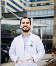
Mehmet Emin Adin, MD
Faculty Member
Yale University, Department of Radiology and Biomedical Imaging
© 2026 Medality. All Rights Reserved.
