Case Review Live - Adult Suprahyoid Neck, Dr. Gloria J. Guzmán Pérez-Carrillo and Dr. Rami Eldaya (10-12-23)
HIDEInteractive Transcript
0:02
Hello and welcome to Noon Conference,
0:04
hosted by M R I Online Noon Conference connects the global radiology
0:09
community through free live educational webinars that are accessible for all
0:14
and is an opportunity to learn alongside top radiologists from around the world.
0:19
We encourage you to ask questions and share ideas to help the community learn
0:23
and grow. Today we're honored to welcome Dr.
0:26
Gloria Guzman Perez Creo, and Dr.
0:29
Rami Alday for a live case review entitled Adult Super OID Neck.
0:35
Dr. Guzman completed her radiology residency at West Virginia University and her
0:39
neuroradiology Fellowship and Research Fellowship at the Mallinckrodt Institute
0:43
of Radiology at Washington University in St. Louis,
0:47
where she's currently associate professor of radiology.
0:50
She has special research interest in advancing neuroimaging techniques,
0:54
including advanced diffusion imaging for head and neck cancer diffusion spectrum
0:59
based imaging and molecular imaging of brain tumors with FDO ppa in the
1:03
evaluation of I D H wild-type glioblastoma in additions to outcomes and
1:08
translational research of M R I I M R I in the field of neuroradiology imaging.
1:14
Dr. Alday completed residency at U T M B and Fellowship in neuroradiology at
1:18
Mallinckrodt Institute of Radiology.
1:21
He specializes in neuroradiology cancer interpretation in the brain, spine,
1:25
and head and neck region,
1:26
and is currently an assistant professor at MD Anderson Cancer Center.
1:31
At the end of the case review,
1:32
please join them in a live q and a session where they will address questions you
1:36
may have on today's topic.
1:38
Please remember to use the q and a feature to submit your questions so we can
1:41
get to as many as we can before our time is up. With that,
1:45
we're ready to begin today's case review. Dr. Guzman, Dr. Alday,
1:48
please take it from here.
1:51
Hi everyone. Uh, thank you so much for joining us today. Um,
1:55
and thank you Ashley, for such a nice introduction.
1:58
So we're just gonna get right to it. Um, so a little bit about the format.
2:02
We're gonna show you a, um, indicative image,
2:07
then we're gonna preview the audience question, uh, show you the case,
2:11
go through a little bit of didactics,
2:13
and then go to an audience poll for the question.
2:17
So the first case we're gonna review is Dr. Gio left node Noter.
2:22
Um, so this is the indicative lesion. Um, and,
2:27
um, think about it and we'll discuss it here shortly.
2:30
And this is the audience question that I want you all to think about, um,
2:34
as we do the presentation. So, node sub displays the longest choline muscle,
2:39
how A, there's no displacement, B anteriorly, C posteriorly,
2:43
or d I don't know. So, um,
2:48
let me show the image. So this is a patient that came to us, um,
2:53
with, uh, difficulty swallowing and some, um, dysphagia.
2:57
And you can see that there is this, uh,
2:59
reen enhancing lesions centrally necrotic, uh,
3:03
located at the lateral retropharyngeal space and extending into the, uh,
3:07
per pharyngeal space. You can see the normal pharyngeal space on, uh,
3:11
the contralateral sides here, and you can see how small it is on the other side.
3:15
There is mass effect, uh,
3:17
on the mucosal space and the oropharynx here. Um,
3:22
and this is a tors to baris.
3:24
This is a fosil or you can see how there's mass effect from that lesion as well.
3:30
Now the is, and um, I just have the component or the associated, uh,
3:35
pet CT image, uh, for you to see that it was, uh, abnormally,
3:39
metabolically active. Right? Um, so the reason I wanted to,
3:44
um, discuss this area of the, um,
3:48
of the, um, super hyoid neck, um,
3:51
is that it is an area that is very difficult for the clinicians to see.
3:56
So just a little bit of history on the name of the not revia.
3:59
So Henry Revia was an atomic, uh, anatomy professor in France. Um,
4:04
he actually wrote a very extensive book on, uh, human lymphatic system.
4:07
And all the lymphatic levels that we utilize today actually arises from this
4:12
work, and it is due to him that we give it this name.
4:17
So the nodal ruber is located in the lateral, uh, retropharyngeal space,
4:22
and it is located anterior to the long colline muscle.
4:25
They extend from the C one C two level to the level the OID bone.
4:29
As you can see, um, usually the size is very, very small, three to five,
4:34
uh, millimeters. And I don't know if you call that we measured that, um,
4:38
nodal vie air, and it was 2.7 centimeters. So clearly very, very abnormal.
4:44
Um, the reason this is so important to, uh,
4:47
keep in mind when you are evaluating head and neck tumors in the super hyoid
4:52
neck is that these notes are absolutely not detectable,
4:56
not palpable on clinical evaluation,
4:58
even when they're enlarged as the patient that I just showed you. And, um,
5:03
if the clinician doesn't know about it,
5:04
they cannot treat it and it can be a source, um, o of course,
5:09
of residual tumor, uh, resulting in much worse prognosis for the patient,
5:15
um, uh, which actually has been proven in the literature.
5:18
So if we don't tell the clinicians that this notice there,
5:22
they're not gonna treat it, and the, uh,
5:23
patient outcome is gonna be much poorer. Um,
5:26
so please make sure that you look at these, uh, notes in your evaluation.
5:31
So just a brief, um, anatomy review. So, um,
5:35
this is an m r i neck axial T two weighted sequence.
5:40
We can see that the feral space is located posterior to the, um,
5:45
pharynx is here located in this, uh, blue line,
5:49
our longest coline muscles, um,
5:51
which are located posterior to the retro pharyngeal space. And then we have, uh,
5:55
this fat line here and tear to the vertebral body, which is the, um,
6:01
prevertebral space.
6:02
And then lateral to the monga colline muscle is where we have the lateral retro
6:06
pharyngeal space where the nose, uh, live. So I want you to,
6:11
uh, see that if you have, um, a big mass here, right,
6:15
located in the lateral pharyngeal space, that's gonna move the, um,
6:18
longus coline muscles, uh, posteriorly. And that's an indication that,
6:23
uh, you know, also you have a lesion in this space. Now,
6:28
um, this has nothing to do with the no of reve,
6:31
but the longus coline muscles are very important in this region of the neck.
6:35
If they're, uh, uh, you know, displaced posteriorly,
6:38
then you must have a mass anteriorly in the retropharyngeal space and the
6:42
muscles are moved anteriorly,
6:44
then you must have a lesion in the vert row space. Um,
6:49
so the longest colline muscles are a great anatomical marker for location of
6:54
lesions within, uh, the neck, uh, deep spaces.
6:59
So the retropharyngeal space, um, as I mentioned already,
7:04
you must evaluate the longest colline muscles to know, uh, where the, um,
7:08
you know, lesions are located. Uh,
7:11
know that the retropharyngeal space spans from the base of the skull base to the
7:16
mediastinum. Um, as we have mentioned already,
7:20
it is anterior to the vertebral muscles and posterior to the phn and esophagus.
7:24
And this is also important to understand that it's not a single space,
7:27
it's actually a double space with two components,
7:31
the true retropharyngeal space and the danger space.
7:34
And so this is what we're talking about. Okay,
7:36
so you have the anterior retropharyngeal space, which terminates at C seven.
7:41
Uh, this space will not extend into the mediastinum and will not result in, um,
7:47
mediastinitis, whereas danger space is posterior to the, uh,
7:51
retropharyngeal space. Allah,
7:53
it extends past the C seven vertebral body into the mediastinum and can
7:58
cause, uh, severe mediastinitis and other, uh, problems. So this is also a, um,
8:03
source of spread of, um, metastatic disease. Okay, Ashley,
8:07
you wanna run the, uh, audience response. So notice the er,
8:11
what happens with the long longest colline muscles.
8:17
Okay, so let's show the audience response. Okay, good.
8:22
So most of you got the, uh, correct answer,
8:24
which is that they're displaced posteriorly. Okay.
8:29
And then I'm gonna stop sharing. So Dr. Alday can, um, start with case two.
8:35
So we'll turn our attention to the carotid space, uh, next. Uh, and, uh,
8:39
we'll start by showing a representative image of a lesion within the carotid
8:42
space. So you can start thinking about it in, uh, in the meantime. Uh,
8:47
the question is, which, uh,
8:49
which of the following cranial nerves is not considered part of the supra oid
8:53
carotid space? Creon, F 7, 9, 10, 11, or 12?
8:59
So, as I look at the carotid space, the way I like to think about it is,
9:03
and in general and head and neck lesions is placing the lesion in a space and
9:08
then looking on m r i on the signal vascularity and what it's doing to the
9:12
surrounding structures. So when I look at this image here, what I see is, uh,
9:17
a lesion situated in the carotid space that is T two hyperintense,
9:21
and I'm seeing that lesion displacing the, uh,
9:25
internal carotid artery anteriorly. And on T two,
9:28
I do see a lot of what looks like flow voids, uh,
9:31
or T two hypo intensity within the lesion. So
9:39
I'm gonna, uh, ask you a couple of questions here. One is,
9:42
what do you think is the leading differential diagnosis to, to think of? And,
9:46
uh, number two, how many lesions do you guys see on the M R I? Uh, and then you,
9:50
you can write that in the chat if you, if you'd like to,
9:56
I can show you the pet right now. Uh, and as you know,
10:00
we're looking at the pet, the one,
10:02
the first question I want you to think of is what radiotracer we're using here,
10:06
and how can I tell that? And then, uh,
10:08
obviously the other question is how many lesions are present?
10:13
So one thing, you know, when I look at radio tracers, and this is, uh,
10:17
dotatate or somatostatin, uh, receptor,
10:20
which is predominantly used for neuroendocrine tumors,
10:23
including this entity here to differentiate between it and between another
10:26
common entity in the carotid space, normally physiological uptake. Uh,
10:31
and the reason why I can tell that it is,
10:32
is very strong in the pituitary and a variable degree of uptake in the carotid
10:37
or ciliary tissues. And the thyroid, which tend to be generally moderate,
10:41
but can be intense. And I see a very intense uptake in two areas.
10:45
And this is the, uh, uh, the bilateral carotid spaces.
10:48
And this is the benefit of using, uh, the dotatate pet.
10:52
'cause the smaller lesions sometimes can be challenging on M R I. And as,
10:56
as you see here, it might be hard to see on the T two. And even when I, uh,
11:00
look at the post contrast,
11:02
the con lesion might be hard to pick and can be easily missed. So the, uh,
11:06
PET gave give us the benefit of, uh,
11:08
detecting those lesions in addition to allowing us to differentiate between it
11:12
and other common entities, which we'll discuss shortly. The other thing I, uh,
11:17
always wanna discuss with the carotid space is location, uh,
11:21
can help us sometimes predict not only the pathology,
11:25
but potentially which, uh, component of the carotid space. So again,
11:29
as I'm looking here, I can see if significantly artily enhancing lesion,
11:34
uh, with respect to the muscles.
11:36
And I see the internal carotid artery being displaced anteriorly on the
11:40
contralateral side at the bifurcation of the carotid,
11:43
I see a lesion sitting and, uh,
11:45
not quite splaying the internal and external carotid arteries, but, uh,
11:49
given its size, but it's sitting there at the bifurcation,
11:52
and you can appreciate that very nicely on the sagittal images
11:58
with the bifurcation. Okay, so turning our attention into the, uh,
12:03
dis discussion of the, uh, carotid space before we move on.
12:07
So it's always important to talk about anatomy and boundaries when we're coming
12:10
up with a differential diagnosis.
12:12
So the carotid space itself is a cylindrical space that extends from the jugular
12:16
foramen to the thoracic inlet,
12:18
and it's divided superior and inferiorly by the hyoid bone.
12:22
And today we're turning our attention to the supra hyoid carotid.
12:25
Its contents are in the supra hyoid neck that the crown jewel of it is the
12:30
internal carotid artery. Uh,
12:32
and it's located medial and slightly more anterior to the internal jugular vein.
12:37
Uh, and those are the two prominent vascular lesions.
12:40
And it contains four cranial nerves in the supra hyoid neck. Uh,
12:44
there is variability of location of the nerves based on anatomical cad
12:49
resections, but this is the most common appearance.
12:51
Cranial nerve nine typically is anterior situated between the two vessels.
12:56
Cranial nerve 12 is typically medial posterior to the carotid, and, uh,
13:00
10 and 11 are more posterior with a variable location.
13:03
And then along the posterior sheet there is the sympathetic trunk and
13:08
anteriorly there is the, an cervical, which, uh,
13:12
forms from C one C two and C two C three nerves and supplies the infra hyoid,
13:17
uh, mus, uh, infra hyoid muscles. So based on the internal contents, you know,
13:22
hence the differential for lesions can be formed, uh, with, with, uh,
13:26
with respect to the, the space. So the, so the margins anteriorly, uh,
13:31
anterior to the carotid, uh,
13:33
there is masticator and per pharyngeal space medially,
13:37
there is the retropharyngeal space that Dr. Guzman discussed laterally.
13:41
There's a parus space, which we'll discuss subsequently and posteriorly,
13:44
there is the paravertebral muscles, uh, and their, uh,
13:49
their com uh, components. So speaking about the differential diagnosis,
13:54
uh, this lesion is, uh, paraganglioma. And, uh,
13:57
talking a little bit about paragangliomas of the head and neck,
14:00
they're typically rare entities of tumors in the head and neck and constitute,
14:04
uh, a little bit more than 0.5%. Now, unlike a lot of other areas in the body,
14:09
they're predominantly parasympathetic. 'cause as you know,
14:11
paragangliomas can be sympathetic and parasympathetic.
14:13
The sympathetic typically secrete the catacholamines.
14:16
The parasympathetic typically do not, and these typically do not secrete, uh,
14:21
the most common of them is the carotid body tumor constitute about 60%.
14:25
In 25% of the cases such as this case, they can be multicenter.
14:29
And the majority of the time, if they are,
14:31
they're typically related to a syndrome or familial. Um,
14:35
one important thing to understand when it comes to syndromes,
14:38
the ate dehydrogenase mutation is at the center of the majority of these
14:42
syndromes, and there's multitude of genes that contribute to it.
14:45
And there's multitude of paraganglioma syndromes associated with this.
14:48
I sh the case that I showed you right now is a, uh, paraganglioma,
14:54
uh, syndrome, familial syndrome one, and then one in three, for example,
14:58
have a high propensity to be present the paragangliomas in the neck.
15:01
Another syndrome that's associated with it is carish triad,
15:05
where you have just tumors, uh,
15:07
lung chos and you have paragangliomas. And in fact, you know,
15:11
I've seen one last couple of weeks, m e m type two NF and, uh,
15:15
one and von Hippa Linda are other things.
15:17
So when you see multiple paragangliomas of the head and neck,
15:20
those are things you want to think of in terms of, of imaging. Again, as I,
15:24
you know,
15:24
going back to the anatomy location and help you predict which nerve it is.
15:29
So the carotid body tumor is the one that displays the I E C A
15:34
and i c a and sits in between 'em as, as I showed you in that image.
15:38
The Glo panicum, uh, tumor, uh, tumors are, you know,
15:42
sit at the cochlear promontory and we're not talking about them. Uh,
15:45
today at the skull base arise from cranial nerve nine.
15:49
And then the jugular typa,
15:51
or more jugular in the jugular vein arises from cranial nerve 10.
15:54
And CLOs vali are the ones that most commonly are located between the jugular
15:59
foramen, you know, and the supra hyoid neck.
16:00
They have propensity to be located at the lateral ma uh,
16:05
mass of C one,
16:06
because the majority of them arises from ganglion nado at that level.
16:10
And the case I showed you is one of those. Uh, and then given their location,
16:14
when you think about it, cranial nerve 10, where it's located,
16:17
as we talk about it here,
16:19
that location is gonna push the vessel 'cause it sits behind the internal
16:24
carotid artery, is gonna push it anteriorly.
16:26
So that's one clue for you to determine the origin or which nerve is, is,
16:31
is the responsible for para paraganglioma. And lastly,
16:34
in especially in familial symptoms,
16:36
you can have laryngeal paragangliomas that arise from the laryngeal per ganglia.
16:39
And in there, typically located in the visceral space,
16:42
similar to other paragangliomas,
16:44
they're arterially enhancing and they do not look like mucosal lesions.
16:48
They're typically submucosal. And you know, so they're, they're,
16:51
you are looking at them, you're like, it doesn't look like a carcinoma.
16:54
So you see an arterially enhancing lesion in, you know,
16:56
the larynx that is not related to the mucosa.
16:59
One of the things to think about is definitely paragangliomas and syndromic
17:03
paragangliomas. In fact, I've seen one last week.
17:06
So very quickly in terms of imaging on ultrasound,
17:09
this is ultrasound they did a couple of days ago for a different patient. Uh,
17:13
they tend to be hypo coic. Again,
17:15
they're very intimate relationship with the vessels given their, uh,
17:19
location in the carotid space. And, uh, they are very vascular,
17:24
such as this case. This is the ultrasound, and this is the companion ct.
17:28
You can see the internal external, uh, carotid arteries. You can see, uh,
17:31
a lesion displaying them and significantly vascular.
17:34
And this is its appearance on the ct, on sagittal images on m r I,
17:39
they're typically T two hyperintense, the flow voids on T two.
17:42
And then on T one you can have the hyperintensity, which is reflective of,
17:46
you know, slow flow within the vessels or, uh, uh, turbulent flow. And it's, uh,
17:51
two things that, uh, I wanna emphasize is one,
17:54
the significance of of not doing a biopsy on these, uh,
17:59
'cause we perform biopsies.
18:00
So not doing a biopsy and being mindful of thinking about that differential and
18:04
angiogram for treatment planning. Uh,
18:06
most common differential would be a nerve sheet tumor and give,
18:10
they're typically given, uh, they're, they're arising from, you know, you know,
18:14
the nerves they're displayed, they tend to displace the, uh,
18:17
vessels anteriorly or, uh, medially the carotid. And then additional, uh,
18:21
lesions always because of the vessels, the vascular,
18:23
and there's a whole gamut of it that's hard for us to go through. Uh,
18:27
which include dissection, FERBs aneurysm, pseudo aneurysm, carotid blowout,
18:31
F M D, vasculitis, uh, and then nia or face syndrome or typic syndrome,
18:36
multiple names for it. And then always remember lymph nodes, uh,
18:39
whether it's infection met or lymphoma. Uh,
18:42
a couple of common lesions I wanted to talk very quickly about.
18:45
This is a carotid space, schwannoma. And, uh, things that, uh, you know,
18:50
typical appearance of schwannoma is, you know, typically T two hyperintense,
18:53
but different, uh, appearance based on the content. And as you can see here,
18:57
it's displacing the carotid laterally. Uh, and then on, uh, doted eight pet,
19:01
it doesn't demonstrate increased uptake.
19:03
And that's one way you can differentiate if you're not sure between car, uh,
19:06
schwannomas and paragangliomas. Other thing I want to really, uh,
19:09
talk about very quickly before we, uh, wrap up the car,
19:12
the carotid space is carotid blood. Because, you know, in, in my my practice,
19:17
I do see quite a decent amount of these, so I want to touch base with it.
19:20
'cause I know not a lot of people see him much.
19:22
Carotid blowout is a potential risk for treatment of head and neck cancer,
19:26
seen in about three to 5% of patients that have, you know, uh,
19:30
surgery and up to 10% with people that have radiation. Uh, and then the, uh,
19:35
what, what happens is it, it's,
19:38
there's an effect on the vessel that increases the risk of the vessel to bleed
19:42
and is classified as, uh, threatened where we see abnormal imaging of vessel,
19:47
whether, whether it's, you know, changing in caliber of pseudo aneurysm, uh, or,
19:51
you know, stenosis, uh, or it can be imminent or, you know, uh,
19:55
active where you see active bleeding or blush on your arterial face.
20:00
Things that increase the risk of it is location of tumor with respect to the
20:03
vessel, if it's encasing the vessel, uh,
20:05
narrowing changing caliber of pseudo aneurysm, uh, or, you know,
20:09
obviously active bleeding.
20:10
And one big thing that it seems to be more sensitive is necrotic tissue
20:15
extending toward the vessel.
20:16
This is a patient that had had the neck cancer that was treated with, uh,
20:19
radiation and surgery,
20:22
and you can see the necrotic tissue and ulceration from radiation.
20:25
And some of the E C A branches, you can see here the caliber of the vessels.
20:29
So I'm going Coggle to cranial, there's significant narrowing of two vessels,
20:33
two branches, this is a.here, and then that increases its caliber,
20:37
and then it's, you know, that's the normal caliber of the vessels.
20:40
You can see that change, uh, change in caliber. This patient came in with, uh,
20:44
uh, active bleeding at the time, time, uh, and had to have, uh, a, uh,
20:49
a stent placement. So that's, uh, pretty much, you know,
20:52
the carotid space and some of its differential diagnosis.
20:55
And then going back to the audience question, Ashley,
20:58
if you wanna ask us the response?
21:03
Yep, Dr. Yep. Perfectly, uh, correct. Seems like everyone you know was,
21:07
was on board on that. Yep.
21:11
So case three, um,
21:13
is the tibular trigone extending to the perennial space.
21:18
So this is a key image. Um,
21:20
and you can see that there's a large mass here on the right side causing
21:25
mass effect on the, um, oropharynx.
21:28
So the audience question for you guys to ponder while we do the presentation,
21:33
displacement of the peripheral andal, uh,
21:35
fat medially and posteriorly means the primary tumor originates in which space?
21:39
A masticator space B parotid space C uh, pharyngeal mucosa,
21:44
dec carotid space, or E um, I don't know.
21:49
So the, um, retro mandibular trigone is actually part of the oropharynx.
21:54
However, it can have extension into the peripheral NAL space,
21:57
which is located in the supra hyoid, um, neck.
22:01
And this is an MR image with normal anatomy.
22:04
So the retro mandibular trigone in parallel to the, uh,
22:09
nodal ruber cannot necessarily be palpated by the, uh,
22:14
E N T clinician or the referring clinician. Um, it is located in this, uh,
22:19
little space of fat here, um, between, uh, the, uh,
22:23
teeth and the body of the mandible. Uh,
22:27
this is the base of tongue here, and these are domesticated muscles. And again,
22:31
it's very important, uh, for us to look at this region in our anatomical images,
22:36
because if we don't talk about it, uh, lesions in this region can, uh,
22:40
go undiagnosed, uh, which causes, uh, poor prognosis.
22:47
So, uh, the retro mandibular, uh, trigone, again,
22:50
is a subside of the oral cavity, um,
22:53
which consists of the mucosa posterior to the last mandibular molar. Um,
22:57
because of its, its location,
22:59
it has a propensity to extend into the peripheral andal space,
23:02
which is part of the super hyoid neck. And like I just mentioned already,
23:07
if you take nothing else of this, of this entire presentation today, study,
23:12
you know,
23:12
retropharyngeal lymph nodes and the retro mandibular trigone cannot be palpated
23:17
appropriately by the clinician. And if we don't mention it,
23:21
it's gonna go undiagnosed, uh, by the referring clinician, and of course,
23:25
result in much worse prognosis for the patient.
23:30
So, um, again, a brief review of anatomy. Again, this is an Mr.
23:35
Axial image of the neck at the level of the, um,
23:39
retro mandibular ular trigone and the paranal space. Um,
23:45
T two weighted imaging, which we can tell by the bright C s F.
23:48
So the masticator space, uh, here in red, um,
23:51
contains the masticator muscles, um, uh, the masseter muscles,
23:56
the regrade muscles, and the temporalis muscles. Of course,
23:58
we have the parotid space, which contains the parotid mass.
24:02
We have the mucosal space here. Again,
24:05
the longus colline muscle is such an important, uh,
24:08
imaging anatomical marker for us in the head and neck. Um,
24:11
so try to become familiar with it and lateral to the longus colline muscle and
24:16
medial to the ator spaces where we have the paranal space. Uh,
24:21
similar to the carotid space, it is, um, separated, uh, in, uh,
24:26
by the styloid processing to the pre, uh, uh,
24:29
styloid peral space and the post, uh, styloid, uh, peral space.
24:35
So the contents of the per Ange space will be discussed in the next case by Dr.
24:40
Aya.
24:41
It is important to understand what the boundaries of the paral space are.
24:46
So again, as I mentioned before, uh,
24:48
laterally we ha laterally and anteriorly. We have domesticate space,
24:53
uh, laterally and posteriorly.
24:55
We have the deep lobe of the paric land with the, uh,
24:59
retro mandibular vein, uh, located here, uh, posteriorly.
25:04
We have the, uh, styloid, uh, process as well as the, uh,
25:09
tensor molecular styloid fascia, which is this, uh, pipeline that, uh,
25:14
we see here. Um, and anteriorly, we have, of course, the, um,
25:19
uh, uh, the tego mandibular rae, uh, that extends from the medial,
25:24
uh, tego plate, um, to the mylohyoid line. Now,
25:28
why is it important to understand, um, all of these, um,
25:33
kind of boundaries for the per pharyngeal space? So,
25:35
similar to the longest colline muscle,
25:38
depending on where the tumor is located,
25:42
that perineal space fat is gonna be displaced.
25:46
So if you have, uh, pharyngeal mucosal lesion,
25:50
which is located anterior and medial to the perge space,
25:54
that per pharyngeal fat is gonna be displaced posteriorly and
25:59
laterally. If you have a masticator, um,
26:03
space mass,
26:04
which is located anterior and lateral to the per pharyngeal space,
26:08
you're gonna have medial and posterior displacement of the per
26:13
a**l, um, fat. If you have a mass in the parotid space,
26:18
which again we mentioned is in the lateral and posterior aspect of the per
26:22
pharyngeal space,
26:23
that's gonna move your peripheral a**l fat anteriorly and medially.
26:30
And finally, if you have a carotid space tumor, which is located, uh,
26:34
posterior and medial to the peral, uh, fat,
26:38
that peral space is gonna be displaced anteriorly and
26:43
laterally, just like the longest colline muscles.
26:46
Understanding this anatomical relation between the peroneal space and
26:51
the spaces surrounded,
26:53
it is critical to understand where the iso center of your lesion is.
26:58
Uh, and I highly encourage you, uh, to all to practice, um,
27:03
locating masses by looking at the displacement of the peroneal, uh, uh,
27:08
space fat. Okay, Ashley, so audience response now,
27:13
displacement of the peroneal fat mely and means the tumor
27:18
originates in which space.
27:24
Okay, great. So most of you got the correct space, which is masticator space.
27:28
Perfect. Okay, on to the next case then.
27:33
Okay. So moving on. Uh, we, uh,
27:36
this is a nice segue to the per pharyngeal space representative image. Uh,
27:40
m r i facet T two. And our, uh, audio question is,
27:45
uh, which of the following is not a content of the peral space,
27:49
fat lymph nodes, uh, uh,
27:52
V three branches of the trigeminal nerve or, uh, ciliary tissue. Uh,
27:57
and as we're thinking of this, we'll start discussing the case, uh, and again,
28:00
you know, similar to what Dr. Guzman have said before,
28:04
always location of the epicenter of the mass, and then from there,
28:07
the differential diagnosis, uh, of the lesion.
28:10
So I'm looking at a lesion here that is in the supra hyoid neck,
28:14
and it looks like it's predominantly situated in the per pharyngeal space,
28:17
comparing to the other side where the fat is suppressed and it's not, uh,
28:21
outside of a little bit of mass effect on the, uh, medial intergrade muscle,
28:24
it's not doing a whole lot of displacement.
28:26
So I know more likely than none that the,
28:29
the space is intact and it's most likely coming from the space. Uh, it is very,
28:33
uh, T two hyper intense and on post contrast sequence,
28:37
it demonstrates, uh, significant, uh, enhancement, uh,
28:41
to bits and portions of it while other par parts are not enhancing. Uh,
28:45
and then my lead differential when I see a lesion like this in the per
28:48
pharyngeal space is gonna be either a, uh, salivary, uh,
28:53
gland or salivary tissue, uh, primary lesion or a, uh,
28:57
neurogenic lesion, just knowing what, which, which of the two are,
29:00
are the most common.
29:02
And then based on the imaging appearance and the significant, uh,
29:05
T two hyperintensity hyper, you know, I would favor, uh, something like, uh,
29:09
benign mixed cell tumor or pleomorphic adenoma as the primary lesion would,
29:13
would the back of my mind thinking of something of schwannoma as the less likely
29:17
consideration given the appearance on, on the sequences. So moving forward,
29:22
you know, again, think about the differential. Uh,
29:25
let's talk a little bit about the anatomy, uh, contents and, and,
29:27
and boundaries. And I know Dr. Guzman talked about a little bit of boundaries,
29:30
so we'll, we'll skip that and just talk predominantly about the contents.
29:34
The majority of the spaces made of fat. Uh, it does have, uh,
29:38
to a variable degree, variable vessels from, uh, the external carotid, uh,
29:43
artery branches. Uh, and then from the tego venous plexus, again,
29:47
there's variability of how much vessels, if any,
29:49
are present based on the variable anatomy, but typically more likely than none,
29:53
there is, and there is branches of V three in particular supplying the, uh,
29:57
tensor valley palatini muscle and ciliary tissue.
30:00
And there's always a debate if, if the primary is, uh, ciliary,
30:05
is it truly from a, uh, you know,
30:07
minor ciliary tissue in the periphery andal space,
30:10
or is it exo coming from the deep lobe of the adjacent parotid?
30:14
And sometimes it is really hard to say. Sometimes it might be easy as Dr.
30:18
Uh Guzman said,
30:19
based on the displacement of the space or if there's residual space. Um,
30:23
in terms of the lesions of per perianal, the majority of the lesions are benign,
30:28
uh, 75 to 85%. Uh,
30:30
it does not constitute much of the primary head and neck tumors. As you know,
30:34
0.5% of all of the primary head and neck tumors arise in the per pharyngeal
30:38
space.
30:39
The most common primary of the perianal space is gonna be salivary gland, uh,
30:44
followed by neurogenic, uh, of the salivary.
30:47
The most common is benign mixed cell tumor or pleomorphic adenoma.
30:51
And of the neurogenic, uh, paraganglioma, typically from, uh,
30:55
vagal nerve branches, followed by schwannoma. And then,
30:58
given that the majority of the, uh, of the content is, is fat,
31:02
there's always, you know, a possibility of lipoma or lipos sarcoma.
31:06
And certainly infection spread from adjacent spaces is another consideration.
31:12
So when talking about differential, as we spoke earlier,
31:15
this is a paraganglioma. The reason why I think it's paraganglioma,
31:19
and you can see the contralateral fat,
31:20
predominantly fat and small vessels in the space,
31:24
is that it's, uh, significantly enhancing.
31:27
And I can see increased vascularity along the margins.
31:30
So I would favor paraganglioma schwannomas. You know, we have, uh,
31:35
variable signal,
31:35
but typically they're T two hyperintense and they demonstrate a degree of
31:40
enhancement. Uh, they are usually on CT hypo intense to the muscle.
31:45
So when I'm looking at a lesion like this,
31:47
I am thinking it's not gonna be paraganglioma because it's not significantly
31:50
enhancing on ct. And then on T two,
31:53
the typical appearance of a benign mixed cell tumor is, you know,
31:57
more bright or more T two hyperintense. So I'm thinking, you know,
32:01
it's probably not gonna be an I would lead with schwannoma in, in this instance.
32:05
Uh, and then, you know, every now and then you'll see one of,
32:08
one of these lesions here. This is the perineal space on, on the left,
32:12
and you can see the right, again, predominantly fat.
32:14
And you can see that this lesion, there is expansion, uh, of the,
32:18
of the perineal space. And there is, you know, fat content and heterogeneous,
32:22
uh, tissue inside of it. So this would be, you know, a fat, fat lesion.
32:27
And certainly if you have an M R I, you can confirm it with, with the fat,
32:30
fat sequences. And I would, you know, given that I do see soft tissue,
32:34
I would favor that this would be a little bit more aggressive than lipoma,
32:37
possibly a lipo sarcoma, which was the case on, uh, this case.
32:41
And then this is another case, you know, more common in pediatrics,
32:44
but you tend to occasionally see it in adults.
32:46
This actually case I read last week is a patient, uh,
32:50
30 years old that came in with, you know, uh,
32:52
difficulty breathing and swallowing. And you can see a T two, uh,
32:56
hyper intense mass, predominantly situated in the per pharyngeal space,
33:01
but it does extend to the pharyngeal, uh, walls and compresses the pharyngeal,
33:05
uh, uh, the nasal pharyngeal airway at the same time, you know,
33:09
it is at the margin of the deep lobe of the parotid, but notice this is not,
33:12
you know, uh, doing a lot of mass displacement. It's, you know,
33:16
kind of spacial and embedding into the spaces.
33:19
So if you look a little bit closer, there's also another component in the, uh,
33:23
retro ular space. So when I was reading this with one of our fellows,
33:26
there were, you know, the thought process was, could this be a, uh, uh, pre,
33:30
you know, again, with the differential, could this be a, uh, a, a,
33:33
a schwan or could the, is this be benign mixed cell tumor,
33:37
or could this be pleomorphic, uh, uh, adenoma, or could this be, uh,
33:41
something like a paraganglioma? And then this is the dotatate pet,
33:45
and there's no increased signal. But if you look closely,
33:47
you can see some calcifications in the ocular component or separate lesion.
33:52
And we thought that this is a low flow vascular lesion,
33:55
which ended up being the case. Now, these are more common in,
33:58
in younger patients in the per pharyngeal space,
34:00
but you do see 'em every now and then in the, uh, adults.
34:02
So going back to our audience question to, to, to recap, uh, you know,
34:07
the, the space,
34:08
which of the following is not a content of the per pharyngeal space?
34:14
Okay? And, and then the majority of, uh, if you got it right, yes. Remember,
34:19
you know, there are a few, uh, typically V three branches in particular, uh,
34:24
the, uh, you know, branches, uh, supplying deten vini. And then, and,
34:28
and then hence why you will have nerve, uh, you will have, uh,
34:30
neurogenic tumors like, uh, paraag, gliomas and schwannomas.
34:34
And then the other thing when it comes to, uh, ciliary tissue, again, you know,
34:39
there, you know, potentially debatable,
34:41
is the ciliary or the most common tumor being the benign mixed cell tumor,
34:44
is it arising actually from the perineal space or the deep lobe? And,
34:48
and there is definitely perineal, uh, uh, perineal space,
34:53
manous delivery gland. Typically, there's no lymph nodes in the space. Okay.
34:57
And then with that, moving on to Dr. Guzman,
35:02
Our next case is a parotid mass with perineal spread of disease. Um,
35:07
as hopefully most of you know, uh,
35:09
per capita adeno cystic carcinoma is the tumor with the highest rate of
35:14
peral spread of disease. Although in, um, in brute numbers,
35:19
it is squamous cell carcinoma,
35:20
just because it is so much more common than adeno cystic carcinoma. And, uh,
35:25
these are the, uh, key images. So now I'm going to, uh, go to the case.
35:31
All right, so this is a person that presented with a parotid space mass.
35:36
Um, this is a flare, uh, sequence,
35:39
and you can see that there's significant enlargement of the entire, uh, gland.
35:44
Remember that to differentiate between the superficial and the deep lo the
35:48
parotid, you need to use the retro mandibular vein.
35:51
This is important because the, uh,
35:53
cranial nerve seven runs in the deep space of the parotid lobe.
35:57
And when the surgeon is, uh, operating, they need to know, um,
36:02
if they're gonna have to go into the deep space of the parotid gland as, uh,
36:06
they're at risk of course, of injuring the nerve. As you can see,
36:10
there's significant restricted diffusion along the entire parotid gland,
36:16
uh, which is, uh, very abnormal. Um, 70% of parotid, um,
36:21
mass lesions tend to be benign pleomorphic adenomas. And, um, they don't, uh,
36:26
show this, um, significant increased restriction, um, again,
36:30
accompanied by, uh, low a d c values,
36:33
which is compatible not with T two shine through,
36:36
but with actual true restriction. We can see that there's a lot of, um,
36:41
vascularity in this lesion. Um,
36:44
and then there's cord like and enlargement of the auricular temporal nerve here,
36:48
right? So this is the normal, uh, IC gland, which tends to be fatty,
36:53
somewhat bright, um, both on T one and T two weighted sequences.
36:57
But you can see that there's this kind of cord like, uh,
37:00
lesion along what the course of the auricular temporal nervous, uh, which is,
37:05
uh, clearly abnormal. Then as we move, uh, onto our,
37:10
uh, enhanced images, um,
37:14
we can see the same kind of appearance, right?
37:16
So we have a significantly enlarged, uh, parotid, uh, gland,
37:20
both the superficial and the deep lobe. Again,
37:23
you have to find that retro mandibular vein to separate both.
37:27
And you can see this cord like thickening, uh, uh,
37:31
an abnormality of the auricular temporal nerve, um, which, uh,
37:36
of course is suggestive of, uh,
37:38
auricular temporal nerve invasion and peroneal spread of disease.
37:44
Um, in fact, this patient, um, when they went, uh,
37:48
to pathology, uh, after resection, uh,
37:51
it was confirmed on pathological evaluation that they did have perineural spread
37:56
of disease, and that the, um, tumor was in fact an adeno cystic, uh,
38:01
carcinoma, which, um, was suspected based on the perineural, uh,
38:05
appearance. So, for the audience question, things to think about,
38:10
um, as I do the presentation, um,
38:13
the UROP temporal nerve is in which perineural, uh, spread highway,
38:18
uh, corneal nerve, uh, six to seven, five to seven,
38:23
eight to seven, I don't know.
38:28
So this is critical in the evaluation of perineal spread of
38:32
disease, especially if you're dealing with superficial lesions to the parotid.
38:37
So it doesn't only have to be a parotid mass, it can be, um, like, uh,
38:41
skin squamous, cell carcinoma, um,
38:44
melanoma or metastatic disease from, um, different, uh,
38:49
uh, metastatic tumors that might affect the face, because we have, uh,
38:54
two different large nerves that, uh, live in that area.
38:57
Sorano nerve five and chrono nerve seven.
38:59
And you can see that there are areas where they interact with each other, right,
39:04
where they, um, mix with each other.
39:06
The most common one being this auricular temporal nerve, which is,
39:09
is a branch of, um, which is a branch of V five,
39:14
but then it has a connecting branch to the Corda Symphony,
39:17
which is a branch of V seven, uh, with the Corda Symphony living within the, um,
39:22
the deep space of the IC gland. And this is that, uh,
39:26
highway that we see here, uh, when we see it affected in this patient.
39:32
Um, so, uh, this is another case from the literature. Um,
39:38
this is the, um, mandibular, uh, ramus red and the mandibular condyle.
39:44
The, uh, OT temporal nerve runs in a C shape behind, uh,
39:48
this area and connects to V five, right?
39:52
This is the foramen oval. This is foram noval here on the right,
39:57
on the left side, which is, um,
39:58
abnormally en large and enhancing normal on the right side.
40:02
And you can see that there's this cord like thickening and enhancement compared
40:07
to the normal contralateral side. Remember, again, that,
40:11
this or temporal nerve, then we'll connect to the Corda Symphony, uh,
40:16
branch of cranial nerve seven, living in the deep lo of the parotid,
40:20
causing that, uh, cranial nerve five to cranial nerve seven, uh,
40:24
perineural highway, uh, spread of disease. Um,
40:29
this is another example from the literature. Um, this is the same finding,
40:34
but on the other side, right? So, um,
40:38
this is the Anglo basal fascia, which is around the nasopharynx.
40:42
And lateral to that is where we find our, uh, cranial nerve, uh,
40:46
five for amino valley. So this is abnormal for Amino Valley, not enlarged,
40:50
not enhancing. This is abnormal amino valley,
40:54
very enlarged and enhancing with associated cord like
40:58
thickening and enhancement of the orical temporal nerve. Again,
41:03
very concerning for, uh,
41:05
perineural spread of disease and putting the patient at risk of the cranial
41:09
nerve five, right to cranial nerve seven, uh, per, uh,
41:13
perineural spread of disease highway, uh,
41:15
connecting to the Corda Symphony in the deep lobe of the paric gland.
41:21
Now, there are multiple, uh, cranial nerve, five to seven highways.
41:26
Um, so I'll just mention a couple of the more, uh,
41:31
um, um, more prominent ones.
41:34
So hopefully everybody knows that the, uh,
41:37
gr superficial petrosal nerve arises from genic ganglion,
41:42
um, which is the ganglion of cranial nerve, uh, seven.
41:46
And via that lingual nerve, we can have a, um, uh,
41:51
greater superficial petrosal nerve, uh, uh, highway, uh,
41:55
that connects with the, uh, cranial nerve five. Here again,
42:00
we have an abnormal enlarged from valley, which carries, again,
42:04
cranial nerve five, abnormal enhancement of the ness sinus with, uh,
42:08
retrograde, uh, extension through the greater petrosal nerve, uh,
42:13
into the genetically ganglion. Um,
42:18
this is another one, uh, with the, uh,
42:21
tebo palin ganglion connection at V two, um,
42:25
with connection to V seven through the greater superficial petrosal nerve via
42:29
the, uh, uh, the video nerve.
42:32
So hopefully everybody recognizes the structure as the tego palatine
42:36
fossa usually containing fat and some vessels, some nerves.
42:41
But here you can see it's abnormally enlarged, abnormally thickened, uh,
42:46
noting that of course, in the tego palatine fossa,
42:48
we have the branches of V two.
42:50
Then we have retrograde flow through the foramen rotunda, which is the, uh,
42:55
foramen, uh, that carries, uh, c nerve, uh,
43:00
five V two branches back into the, um,
43:04
cabeno sinus. And we see retrograde extension again through that superficial,
43:09
greater superficial petrosal nerve into our geno ganglion with abnormal
43:14
enhancement of both the typa and the labyrinthine section
43:19
of the, uh, facial nerve, uh, cial nerve seven, uh, again,
43:23
demonstrating another cranial nerve five, cranial nerve seven highway,
43:27
this time through V two branches. Alright,
43:32
so we'll do the audience response now.
43:39
Okay, awesome. I am so pleased to see this. Yes. Um,
43:42
definitely the oroc temporal nerve is the, uh,
43:46
cranial nerve five to seven highway, uh, the most important one, although,
43:49
as I mentioned, there's quite a few of them. And with that,
43:53
I'll turn it over to Dr. Alaya.
43:58
So in continuation with Dr.
43:59
Guzman's discussion of perineural spread masticator expense, uh, lends itself,
44:03
you know, really nicely for the next case. So I'm showing you, uh,
44:07
representative image of, uh, someone's 25 years old, and, uh,
44:12
have a, uh, lesion, uh, situated in masticator space.
44:16
Just want you to think what is your, uh, differential? It's always, you know,
44:19
hard with these lesions specifically to come up with a specific diagnosis, but,
44:23
uh, what is, uh, what is your differential to start thinking of? And then, uh,
44:27
the audience question is, which of the following is not a,
44:30
a masticator space muscle, uh, temporalis muscle medial oid,
44:35
uh, the masseter or the vaccinator muscle?
44:40
Okay, so masticator space anatomy is, to me,
44:43
is one of the more fascinating spaces of the supra high neck for a couple of
44:46
reasons. One, uh, it's typically ignored because the pathology is,
44:51
isn't as common as other spaces. Uh,
44:54
number two is there's so many confusing, uh, phrases and so many,
44:59
uh, di uh,
45:00
diversion between surgeons and radiologists in terms of how we define it.
45:04
So I wanted to spend a little bit of time discussing the anatomy,
45:06
the boundaries, and the subspaces before turning our attention to the,
45:09
to the case.
45:10
So starting first by talking about the contents of the masticator space.
45:13
So predominantly the contents are, you know, muscles of mastication,
45:17
which include the lateral, uh, turid muscle, uh, the medial turid muscle,
45:21
the master muscle, and the temporalis muscle. Uh,
45:25
and then there is a small component of the mandibular, uh, bone,
45:29
which includes the posterior body and the ramus. And we also have, uh,
45:34
V three nerves. So in, in keeping with Dr.
45:36
Guzman discussion branches of V three, including for, uh, you know, the, uh, uh,
45:41
V three branches going toward the inferior alveolar canal. And, uh, lastly,
45:47
uh, we have a few vessels, uh,
45:49
also depending on the variation of the anatomy that are branches of the inferior
45:53
alveolar artery vein and oid venous, uh, plexus in terms of, you know,
45:57
terminology in, in, and then you,
45:59
and how we divide it for radiology standpoint. Historically,
46:03
we've divided the space with respect to the, uh, to the, uh,
46:09
uh, zygomatic arch into supra zygomatic space and, uh, you know,
46:13
infra zygomatic space.
46:14
And the supra zygomatic essentially predominantly contains the temporalis
46:19
muscle,
46:19
which is equivalent to what the surgeons typically call the temporal fossa.
46:23
And to the surgeons.
46:24
The temporal fossa is essentially that superior extension of the ma, uh,
46:28
masada space, or the zygomatic masada space,
46:31
and is further divided surgically into potential spaces of superficial
46:36
temporal fossa between the fascia and the muscle,
46:38
and the deep between the muscle and the temporal bone. In terms of the infra,
46:43
uh, zygomatic space, it's, you know, further divided into spaces or, uh,
46:48
sub spaces in the, in our literature historically as, uh, one of the,
46:52
is the mass, uh,
46:53
mass esoteric or sub esoteric space between the master muscle and the mandible.
46:58
Uh, and then we have the, uh,
47:01
tego mandibular space between the medial tego and the, uh, and,
47:05
and the bone, the mandibular bone. And that, uh, essentially, uh,
47:10
uh, corresponds to, you know, the,
47:12
the surgeons basically calling it the infra zygomatic or the pharyngeal
47:16
component of the, uh, of the masticator space. Lastly,
47:20
and to add to the confusion, you hear people sometimes using the phrase,
47:24
the infra temporal fossa or space,
47:26
which is the part of the masticator space that is immediately inferior to the
47:31
skull base. Uh, and then it extends from the pharyngeal wall toward the muscle.
47:36
So those are, and then it's posterior to the, uh,
47:38
posterior wall of the posterior lateral wall of the maxillary signs.
47:41
So those are different terminologies of the space that you might hear people,
47:45
uh, speak of all the time. With that, you know,
47:48
I wanted to switch the order a little bit here, because I, when I go to,
47:51
to discuss the image, I, I wanted to, uh,
47:54
to have that abide as we're looking at this.
47:56
So what I'm seeing here is I'm seeing a T two hyperintense lesion that is
48:01
extending essentially, uh, from the, uh,
48:04
masseter muscle to the temporalis muscle, or in other words,
48:08
extending from the infra, uh, zygomatic, uh,
48:13
masticator space to the, uh,
48:15
suppress zygomatic masticator space that is demonstrating enhancement. Uh,
48:20
and then when I'm starting to think about this from a differential standpoint,
48:23
I, you know, my first feeling is that this is a lesion that is situated in the,
48:27
um, masticated space. Predominantly it's not extending from outside.
48:31
Then I start to work my differential based on the contents of the and location.
48:36
And for me, the overall appearance makes me want to lean toward, you know,
48:40
a primary sarcoma, this lesion here. Now again,
48:43
telling the subtypes of sarcoma might be challenging, uh,
48:47
because they tend to overlap in imaging, with the exception of few of them,
48:50
obviously, the osseous or the, uh, fat containing, uh, uh, sarcomas.
48:55
So that would be the reasoning why I would think, you know,
48:57
I would lead with the sarcoma here. Certainly mets, if known, you know,
49:00
malignancy, uh, would be there. And then, uh,
49:03
nerve sheet tumor would be less likely. Consideration also on board, on,
49:06
on when, when it, when I see a case like this. So further discussion,
49:11
this end up being a synovial sarcoma. Now,
49:13
head and neck sarcomas are in general rare,
49:16
and they represent 0.1% of the head and neck tumors, uh,
49:20
overall, and they're only three to 10% of the, uh, you know,
49:25
sarcomas in general. Uh, and, and then, uh,
49:29
sarcomas overall are in general where,
49:31
and they constitute 1% of all of the solid malignancies. Now,
49:34
synovial sarcomas do overlap with other sarcomas,
49:36
but they are a little bit less aggressive historically. And then classically,
49:40
they are located predominantly in the extremities.
49:43
The worst synovial is a misnomer, right?
49:45
'cause it's not associated with the synovium of a joint.
49:48
And they're most likely or most commonly affect, you know,
49:51
males in their third to fifth decade within the lower extremities. But again,
49:55
as I said, you know, they can occur in the head and neck, exceedingly rare,
49:58
1.1%. If they are in the head and neck, they typically, you know,
50:03
most common location is the peral space and the hypopharynx followed by the
50:07
masticator space, and they tend to be well circumscribed,
50:11
T two hyperintensity alene enhancing as this case. Now,
50:14
when do I consider sarcomas,
50:16
when I'm looking at head and neck lesions versus carcinoma? Now,
50:19
the predominant malignancies obviously are gonna be carcinomas,
50:23
but location help with the, the, the diagnosis. So in general,
50:27
carcinomas are either gonna be cutaneous, right, or they're gonna be relate.
50:31
And those should be relatively straightforward to diagnose, uh,
50:35
and differentiate with sarcomas, with the exception of angios sarcomas.
50:39
Now in the, uh, and or they're gonna be along the mucosal surface.
50:42
So they tend to be deeper or more, uh,
50:45
central versus the sarcomas in general. The other thing is, uh,
50:50
risk factors. So obviously, you know, with carcinomas,
50:53
we know H P V association or, you know, potentially smoking, which, you know,
50:57
most of the time is, is not typically for most of the sarcomas and age.
51:00
A lot of the sarcomas are in younger adults, you know, not,
51:04
not taking into account H P V, obviously, and then lymph nodes. So one,
51:08
another big thing is if it's a lymphoma or carcinoma,
51:11
they're more likely to go to lymph nodes versus sarcomas.
51:14
Sarcomas can go obviously to lymph node,
51:15
but more likely there will demonstrate a hematogenous spread. So, you know,
51:20
to follow up with that, a question, and, and,
51:22
and you can think of is that another masticator space lesion,
51:25
and I'm looking at this as situated within, you know,
51:27
predominantly the medial TER muscle, you know, uh,
51:30
very homogeneously enhancing T two, hyperintense, well circumscribed,
51:33
and then this, this is sarcoma or carcinoma.
51:36
If I'm looking at it and having to choose between the two. And then I, you know,
51:39
I would definitely lean to sarcoma given the overall appearance and location.
51:43
And this ended up being spindle cell, uh, sarcoma.
51:46
So following up and talking about those spaces and potential spaces, the,
51:50
you know, the masticator space, one of the most common, uh,
51:54
the most common pathologies is infection, spread of infection,
51:57
in particular dental infection. Uh,
52:00
this is a patient that had a cyto nasal malignancy. As you can see,
52:03
there's extensive cyto nasal surgery. And then they, uh, post-radiation,
52:07
they had an ulceration along the mucosa, and then, uh,
52:11
they had a superimposed infection. And if you look closely, you will see that,
52:14
uh, sub mesic meric space. And then even the OID space of the ma uh,
52:20
masticator space, you will see, uh, areas of subperiosteal abscess formation.
52:24
So again, you know, uh, infection is one of the most common in,
52:27
in particular orogenic pathologies of the masticator space and something that we
52:31
see very frequently. Another, uh, another lesion that we typically tend to see,
52:35
and it's a little bit more straightforward, is obviously osteosarcomas, uh,
52:39
such as this one here is arising from the ramus. Uh, and, uh, the,
52:43
this patient previously had a lesion that was resected and you can see a large
52:47
recurrence. And then the osteosarcomas tend to have that, you know,
52:51
peri osteo bone reaction. Occasionally the sunburst not here.
52:54
And then comans triad with the lift of the, uh, uh, the periosteum, again,
52:58
not here in this case. And then they, to a varying degree,
53:01
have a soft tissue component, uh, such as this case here. This was, you know,
53:04
a ma mandibular ator space, uh, osteosarcoma that had occurred and,
53:09
and gone into adjacent, uh, space. So to recap, domesticated space,
53:14
it is a space that is very challenging to see on physical exam.
53:18
So imaging plays a central role in diagnosis.
53:21
So we as radiologists should make ourself familiar with it.
53:25
And then it's really important to differentiate between, uh,
53:27
lesions occurring primarily in the masticator space when it comes to
53:31
differential and lesions is spreading to masticator space.
53:34
'cause recurrent and primary carcinomas can frequently extend, uh,
53:38
remember infection and inflammation is exceedingly common,
53:40
especially odontogenic vascular malformation is a more common, uh,
53:44
presentation in pediatrics.
53:46
And we went across one of those lesions in a different space.
53:49
And then neurogenic arising from the actual, you know, V three segments.
53:53
And then remember as Dr. Uh,
53:55
Guzman showed us some examples of perineural spread masticator space do have V
53:59
three branches, and, you know, uh, spread from, uh,
54:02
primary head and neck malignancies is not uncommon. And lastly, you know,
54:06
demandable is part of the space. So pathologies of osseous,
54:09
lesions of demandable are things to think of when you see a masticator space
54:13
lesion. With that, you know, going back to the question,
54:17
which of the following is not a masticator space muscle?
54:24
Yep, absolutely. You know,
54:26
and then we just talked about the temporalis muscle and, you know,
54:29
being part of the supra zygomatic masticator space and then the medial Reid
54:34
muscle, we, we discussed, uh, as, you know,
54:36
part of the masticator of the infra temporalis. And again, you know,
54:39
making yourself familiar with all of these terminologies that vary from a person
54:43
to person and and the literature in between surgeons and radiologists. Okay.
54:47
And with that, I think this is our last case. Uh,
54:50
I'm happy to stick around and answer any questions if anyone has any questions.
54:55
Thank you so much, Dr. Guzman and Dr. Alday. Yes, at this time,
54:58
we'll open the floor for any questions from our audience.
55:01
You can submit your questions through that q and a feature in Zoom,
55:06
so we can get to as many as we can before we have to close.
55:11
Well, I would mention that, um, if any of you have any questions and uh,
55:16
you would like to email either myself or Dr. Alday, please feel free to do so.
55:21
We'd be certainly happy to answer any questions for you. Uh, offline,
55:27
We have one that may have just come through.
55:30
Are there any good articles to refer for neural spread of tumors?
55:35
Um, there actually are, but um, it, uh,
55:39
there's better than that is a, um, a book by Dr.
55:44
Hornberger, um, uh, that,
55:48
that is a head and neck, uh, tumor. Um, uh,
55:52
let me see if I can find, it's a, um, title here
55:57
where it's a case based type of book. It's called, um,
56:02
diagnostic Imaging and Head and Neck. Um, and again, it is, um,
56:06
a book that is, um, edited by Dr. Harberger.
56:11
It's one of the, um, amus, um, volumes,
56:15
and they have excellent, um, case-based, um, anatomy,
56:19
explanation of all the abnormalities of the head and neck,
56:23
including the supra hyoid space, as well as, uh, excellent, um,
56:27
differential diagnosis with, um,
56:29
images of every differential diagnosis as well. Um, so, uh,
56:34
it is a little bit pricey. It's almost $250, but if you can get it, that would,
56:38
it's a great resource.
56:40
What is the most common metastatic to you for the masticator space?
56:45
Rammi, do you wanna take that one?
56:47
Absolutely. So I, I will add to Dr. Guzman that, that the doc,
56:50
the Bookshare is fantastic. The other book I'll say is, is Dr.
56:54
Somms Peter Somms Encyclopedia, but that's, they have,
56:57
he has a very large chapter on peroneal spread about 60 or 70, uh, pages,
57:01
which is fantastic. But the problem is obviously, I mean, affording it if it's,
57:05
it's not a cheap book, but it's a fantastic, uh, chapter.
57:10
And then the other thing, there's a few articles by Dr. Uh, Lawrence Ginsburg,
57:14
uh, discussing, you know, more rare perineural spread in different, uh, uh,
57:18
areas in different branches in the head and neck, which is,
57:20
which are also very fantastic now regarding the, uh, uh, ator space.
57:24
So look at it into two different components. If I'm looking at osseous,
57:28
and it's not metastatic,
57:29
but something that is very common and I see on a weekly basis is multiple
57:33
myeloma and prostate going to the mandible. And in matter of fact, you know,
57:36
not uncommonly, we see people coming in with numbness of the face that have,
57:41
you know, multiple myeloma and they have, you know,
57:43
significant tumor burden involving that inferior alveolar nerve and, and,
57:47
you know, the mandibular canal. Uh, so prost, I'd say,
57:50
if you're looking at the mandibular itself,
57:52
if you're looking at the muscle itself, it's very variable.
57:55
But one of the things that I see more common, and don't quote me on this,
57:58
this is my experience, but not the literature, I don't know of the literature,
58:01
is renal cell carcinoma. I've seen decent number of cases,
58:05
but I don't know exactly, you know, if someone looked at it in the literature,
58:09
I'm not aware of that data. Dr. Dr. Guzman, are you aware of any of that data
58:13
For metastatic disease to domestic cancer? Me,
58:16
Outside of Ossis and, you know,
58:20
No, no, I, I wouldn't know.
58:22
Yeah, I would say definitely Ossis would be number one. And you, you know,
58:25
prostate and, and, and then myeloma not being, you know, not being metastatic,
58:29
but more, you know, intrinsic and then renal from my experience. But definitely,
58:33
you know, a lot of other things.
58:36
Good. And then for the next question, so, you know, again,
58:41
70% of carotid tumors are benign, plu, amorphic, adenoma.
58:47
Um, so when you do see, um, um, orlu, uh,
58:51
or perineal spread of disease,
58:52
then you have to be concerned about more aggressive tumors.
58:57
And cystic carcinoma has to be included in differe differentials or other
59:01
things. Again,
59:01
that can give you peroneal spread of disease or SMM cell carcinoma. Um,
59:06
certainly melanoma, other metastatic diseases less commonly.
59:10
So I would turn the question around and say that when you do see urals spur
59:15
disease, you must always include adeno cystic carcinoma differential,
59:19
although of course, you know, it could be other things as well.
59:23
And I would agree with Dr. Guzman. I will only also add,
59:26
remember that these lesions can skip and the perinatal spread, right?
59:30
So you guys want to, uh, follow the whole course of the nerve before you, uh,
59:36
you make the, uh, you know, assess all of the nerve before, you know, just,
59:39
you know, closing the case and saying, okay,
59:41
there's peroneal spread here or there. Just, you know,
59:43
make sure you follow all of the nerve. Of course,
59:45
especially with endo cystic carcinoma, they tend to, to uh, spread.
59:50
Okay. And then regarding the, uh, boundaries of the, uh, retro mota, I think Dr.
59:54
Guzman had to, to catch your patient. So, uh, you know, the, the boundaries,
59:59
essentially, if you look anteriorly, it'll be, uh, the,
60:04
uh, you know, uh, buccal space posteriorly,
60:07
you are talking about peripheral gene in masticator space. Uh,
60:12
and then one thing I will say, understanding re molar, uh,
60:15
trig is outside of it being a hidden space,
60:18
once you ex reach that area, the, uh,
60:22
spread of tumor just becomes endless. So it, and it's very hidden. Also,
60:26
it's very challenge for us on cts because of the dental amalgam, right?
60:29
So make sure, you know, you get angled views if you,
60:32
if you read a large volume of tumors.
60:34
'cause sometimes you will miss tumors with, uh,
60:37
based on just regular axial through the, uh, the, the, the neck area.
60:42
And then how do carcinoma and sarcoma differ with respect to enhancement and
60:45
bone destruction in general? You know,
60:49
these sarcomas are a whole gamut of, of different, uh, aggressiveness.
60:53
So some of them will, will enhance very significantly. Some of them will not,
60:57
but most of them do tend to enhance pretty strongly. Uh,
61:01
and then they tend to, uh, uh,
61:04
destroy or remodel bone depending on how aggressive they are. So like some,
61:08
something like synovial sarcoma,
61:10
like this sarcoma tend to first push or remodeling before it destroys.
61:14
Similar with, with, you know, spindle cell, but there are, you know,
61:17
sarcomas that are a little bit more aggressive. Um, and then, uh,
61:21
when it comes to, uh, carcinomas, now we do know a lot of, you know, mucosal,
61:25
gingival mucosal and buccal mucosal tend to vary early on, you know, uh,
61:29
invade that mandible, whether, you know, minimal or significant destruction. So,
61:34
you know, in advanced disease or T four disease, when we stage these, uh,
61:38
a lot of times you'll see very frequently oste destruction with carcinomas and
61:42
then the enhancement. You know,
61:44
I would say that it is really hard to just use an enhancement on its own to
61:48
differentiate between the two entities. Again, just because the, the fact that,
61:53
uh, sarcomas are of, of different flavor and different aggressiveness.
61:58
Awesome. Thank you so much for staying on to answer those extra questions Dr.
62:02
Alday. And, uh, thank you so much for the case review today. This was awesome.
62:07
And for our awesome audience for participating in all the questions
62:12
and asking all the questions,
62:15
you can access a recording of today's conference and all our previous noom
62:18
conferences by creating a free M r I online account.
62:21
And be sure to join us next week. We've got two noom conferences.
62:24
We've got one on Tuesday, October 17th, and we're featuring Dr.
62:28
Inez Mohamed for a lecture entitled Psychological Safety as an A C G
62:33
ME Requirement Challenges and Solutions. And then on Thursday,
62:37
October 19th, Dr.
62:39
Steven Rowe is going to join us for a lecture called Current Radiopharmaceutical
62:43
Theranostic Applications in Nuclear Medicine.
62:45
You can register for those lectures@mmrionline.com.
62:48
Follow us on social media for updates on future NOOM conferences. Thank you, Dr.
62:53
Guzman. Thank you Dr. Aya and everyone. Have a great day.

Gloria J. Guzmán Pérez-Carrillo, MD, MPH, MSc
Associate Professor of Radiology, Neuroradiology Section Co-Director, Advanced Neuroimaging Clinical Service
Mallinckrodt Institute of Radiology, Washington University School of Medicine
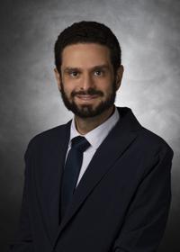
Rami Eldaya, MD, MBA
Assistant Professor
M.D. Anderson Cancer Center
© 2026 Medality. All Rights Reserved.
