Training Collections
Library Memberships
On-demand course library with video lectures, expert case reviews, and more
Fellowship Certificate™ Programs
Practice-focused training programs designed to help you gain experience in a specific subspecialty area.
Ultimate Learning Pass
Unlock access to our full Course Library and all self-paced Fellowships.
Continuing Medical Education (State CME)
Complete all of your state CME requirements in one convenient place.
Noon Conference (Free)
Get access to free live lectures, every week, from top radiologists.
Case of the Week (Free)
Get a free weekly case delivered right to your inbox.
Case Crunch: Rapid Case Review (Free)
Register for free live board reviews.
Dr. Resnick's MSK Conference
Learn directly from the MSK Master himself.
Lower Extremities MRI Conference
Musculoskeletal Imaging
Emergency Imaging
Emergency GI & Abdominal Radiology FellowshipNEW!
PET Imaging
Pediatric Imaging
For Training Programs
Supplement your training program with case-based learning for residents, registrars, fellows, and more.
For Private Practices
Upskill in high growth, advanced imaging areas.
Compliance
NewTrack, fulfill, and report on all your radiologists' credentialing and licensing requirements.
Emergency Call Prep
Prepare trainees to be on call for the emergency department with this specialized training series.
Training Collections
Library Memberships
On-demand course library with video lectures, expert case reviews, and more
Fellowship Certificate™ Programs
Practice-focused training programs designed to help you gain experience in a specific subspecialty area.
Ultimate Learning Pass
Unlock access to our full Course Library and all self-paced Fellowships.
Continuing Medical Education (State CME)
Complete all of your state CME requirements in one convenient place.
Noon Conference (Free)
Get access to free live lectures, every week, from top radiologists.
Case of the Week (Free)
Get a free weekly case delivered right to your inbox.
Case Crunch: Rapid Case Review (Free)
Register for free live board reviews.
Dr. Resnick's MSK Conference
Learn directly from the MSK Master himself.
Lower Extremities MRI Conference
Musculoskeletal Imaging
Emergency Imaging
Emergency GI & Abdominal Radiology FellowshipNEW!
PET Imaging
Pediatric Imaging
For Training Programs
Supplement your training program with case-based learning for residents, registrars, fellows, and more.
For Private Practices
Upskill in high growth, advanced imaging areas.
Compliance
NewTrack, fulfill, and report on all your radiologists' credentialing and licensing requirements.
Emergency Call Prep
Prepare trainees to be on call for the emergency department with this specialized training series.
Lesson
Pre-Course Activities
1 topic, 2 min. of video
Lesson
Introduction to Uterine Imaging and Normal Anatomy
5 topics, 14 min. of video
Lesson
Mullerian Duct Anomalies
11 topics, 40 min. of video
Introduction to Mullerian Duct Anomalies (MDA)
5 m.Agenesis
4 m.Unicornuate with Rudimentary Horn
5 m.Didelphys Uterus – Pediatric
5 m.Didelphys Uterus – Adult
4 m.Unknown case – Septate (Complete Septate Uterus)
7 m.Complete Septate MRI (Fibrous Septum)
6 m.Partial Septate on US
3 m.Bicornuate Uterus - Case 1
4 m.Bicornuate Uterus - Case 2
2 m.Mullerian Duct Anomalies (MDA) – Teaching Points
2 m.Lesson
Intrauterine Device (IUD)
8 topics, 21 min. of video
Lesson
Adenomyosis
10 topics, 27 min. of video
Adenomyosis – Introduction
6 m.Adenomyosis Nodules
3 m.Adenomyosis – Cysts in 26 y/o Patient
3 m.Venetian Blind Appearance of Adenomyosis
3 m.Multiple Imaging Findings of Adenomyosis
4 m.Adenomyosis on MRI – Focal Thickening
3 m.Classic Adenomyosis on MRI – T2 Cysts
2 m.Adenomyosis – Problem Solving with MRI
4 m.Adenomyosis – Problem solving MRI with Endometrioma
4 m.Adenomyosis – Teaching Points
2 m.Lesson
Fibroids
7 topics, 23 min. of video
Lesson
Endometrium
11 topics, 25 min. of video
Lesson
Special Considerations – Post Pregnancy
10 topics, 26 min. of video
Introduction- Post Pregnancy Considerations
1 m.Endometritis (After C-Section)
3 m.C- Section Dehiscence
4 m.C-Section Pseudoaneurysm
4 m.Retained Products of Conception (RPOC)
3 m.Retained Products of Conception and Ancillary Findings
5 m.Arteriovenous Malformation (AVM)
5 m.Arteriovenous Malformation (AVM) – Companion Case
3 m.Isthmocele – Three Appearances
3 m.Teaching Points- Post Pregnancy Considerations
1 m.0:01
Our next endometrial case is a 50-year-old
0:03
who came in for right ovarian cyst follow-up.
0:05
She is postmenopausal at 50 years old.
0:08
So we scroll through this sagittal
0:10
image through the uterus, transvaginal.
0:13
There's a lot going on in this patient as well.
0:15
You can see a lot of cystic change.
0:18
Some of this is going to potentially be blood
0:20
vessels, so you have to figure out if you
0:22
were in the uterus or cervix or next to it.
0:25
But we're going to concentrate on
0:25
the endometrium itself right here.
0:27
And we can see this is abnormally thickened.
0:28
She is postmenopausal per history.
0:31
That is about a 12-millimeter
0:32
stripe, which is too thick.
0:34
for postmenopausal and then
0:37
postmenopausal who is not bleeding.
0:39
But beyond that, we also have
0:40
abnormal morphology with all
0:42
of these cystic spaces in here.
0:43
So this needs a biopsy.
0:46
So we should also look at our color
0:48
Doppler ultrasound imaging to see if we
0:49
can see if there's vascularity to it.
0:51
It's not ultimately going
0:52
to change the management.
0:53
This still needs a biopsy, but it can
0:55
be helpful if it's very hypervascular
0:57
diffusely, if it's focally, maybe this
0:59
is a polyp can be helpful to give the
1:03
guide surgeon a little bit of a heads up.
1:05
And you can see in this particular case,
1:07
it is not really that hypervascular,
1:10
little bits of flow here and there.
1:12
She did end up going to get a biopsy,
1:14
of course, because this is abnormal
1:15
in a postmenopausal patient.
1:17
And they got disordered fragments
1:18
of proliferative endometrium.
1:20
This is considered a non-cancerous
1:22
diagnosis, but is a quote unquote
1:24
unusual growth of endometrial cells.
1:27
So in a case like this, she's going to need
1:28
close follow-up to see if this, you know,
1:30
comes back, or if there is a cancer, or if
1:32
it was just an undersampling type of problem.
1:35
So they'll keep watching her, and she'll
1:38
require follow-ups again to make sure
1:39
that she doesn't end up having a cancer.
Unknown – Proliferative Endometrium
HIDEInteractive Transcript
0:01
Our next endometrial case is a 50-year-old
0:03
who came in for right ovarian cyst follow-up.
0:05
She is postmenopausal at 50 years old.
0:08
So we scroll through this sagittal
0:10
image through the uterus, transvaginal.
0:13
There's a lot going on in this patient as well.
0:15
You can see a lot of cystic change.
0:18
Some of this is going to potentially be blood
0:20
vessels, so you have to figure out if you
0:22
were in the uterus or cervix or next to it.
0:25
But we're going to concentrate on
0:25
the endometrium itself right here.
0:27
And we can see this is abnormally thickened.
0:28
She is postmenopausal per history.
0:31
That is about a 12-millimeter
0:32
stripe, which is too thick.
0:34
for postmenopausal and then
0:37
postmenopausal who is not bleeding.
0:39
But beyond that, we also have
0:40
abnormal morphology with all
0:42
of these cystic spaces in here.
0:43
So this needs a biopsy.
0:46
So we should also look at our color
0:48
Doppler ultrasound imaging to see if we
0:49
can see if there's vascularity to it.
0:51
It's not ultimately going
0:52
to change the management.
0:53
This still needs a biopsy, but it can
0:55
be helpful if it's very hypervascular
0:57
diffusely, if it's focally, maybe this
0:59
is a polyp can be helpful to give the
1:03
guide surgeon a little bit of a heads up.
1:05
And you can see in this particular case,
1:07
it is not really that hypervascular,
1:10
little bits of flow here and there.
1:12
She did end up going to get a biopsy,
1:14
of course, because this is abnormal
1:15
in a postmenopausal patient.
1:17
And they got disordered fragments
1:18
of proliferative endometrium.
1:20
This is considered a non-cancerous
1:22
diagnosis, but is a quote unquote
1:24
unusual growth of endometrial cells.
1:27
So in a case like this, she's going to need
1:28
close follow-up to see if this, you know,
1:30
comes back, or if there is a cancer, or if
1:32
it was just an undersampling type of problem.
1:35
So they'll keep watching her, and she'll
1:38
require follow-ups again to make sure
1:39
that she doesn't end up having a cancer.
Report
Faculty
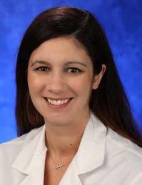
Kathryn McGillen, MD
Assistant Professor of Radiology, Medical Director of Ultrasound
Penn State University Milton S Hershey Medical Center
Tags
Vascular
Uterus
Ultrasound
Neoplastic
Idiopathic
Gynecologic (GYN)
Body
© 2026 Medality. All Rights Reserved.
