Rapid MRI for Acute Pediatric MSK Infection, Dr. Jonathan Samet (7-30-26)
HIDEInteractive Transcript
0:02
Hello, and welcome to Noon Conference hosted by Medallity.
0:06
Noon Conference connects the global radiology community through free live
0:10
educational webinars that are accessible for all, and it's an opportunity to learn
0:14
alongside top radiologists from around the world.
0:18
Today, we are honored to welcome Dr.
0:20
Jonathan Samet for a lecture entitled Rapid MRI for Acute
0:24
Pediatric MSK Infection. Dr. Samet is an associate professor of
0:29
radiology and orthopedic surgery at Northwestern University Feinberg School of
0:34
Medicine, primarily based at Ann and Robert H.
0:37
Lurie Children's Hospital of Chicago, serving as the division head of body
0:42
imaging and section head of MSK imaging.
0:45
He also works in the adult MSK radiology section at Northwestern Memorial
0:49
Hospital. He studies nerve imaging with MRI, neurography, and ultrasound,
0:54
rapid MSK imaging, and MRI techniques for bone marrow evaluation.
0:59
At the end of the lecture, please join Dr.
1:01
Samet in a Q&A session where he will address questions you may have on today's
1:04
topic. Please remember to use the Q&A feature to submit your questions so we can
1:09
get to as many as we can before our time's up.
1:11
With that, we are ready to begin today's lecture. Dr.
1:14
Samet, please take it from here.
1:17
Welcome to the Noon Talk. So today, I'll be speaking about
1:23
rapid MRI in pediatrics for musculoskeletal infection.
1:27
It's a
1:28
technique that we helped develop here at Lurie Children's in Chicago, and we've
1:32
been
1:33
really enjoying it and finding that it really has changed the way that we
1:40
work up musculoskeletal infection.
1:42
It really helps with throughput, and it's been really a game changer for us.
1:47
So I wanted to take you through the
1:50
story of how we did this, and answer some questions of why,
1:56
what, and how.
1:59
And so let's start with why should we do this?
2:02
I want to give a little background of it before we dive into it.
2:06
In pediatric radiology, a little bit different than an adult side,
2:11
we have to deal with problems with sedation and contrast
2:15
use.
2:18
We know that just by giving sedation or general
2:22
anesthesia for a lot of our exams, it can add a significant amount of time and
2:27
strain on the resource in the hospital.
2:29
We do know that there are some risks in pediatric patients of anesthesia.
2:34
And similar in pediatrics and adult, there are long-term risks of
2:38
gadolinium-based contrast use that are unknown.
2:41
But we do know that it deposits in tissues all over the body, so we really should
2:46
try to only use it if we absolutely need to.
2:49
And there are a growing number of indications for non-contrast MRI.
2:54
Every time I talk about this topic, there's one more thing that people are doing
2:58
without contrast. So in musculoskeletal, we have sacroiliitis.
3:04
We're doing an MRI for ovarian torsion now, MRI for
3:07
appendicitis. There's papers on inflammatory bowel disease.
3:12
So
3:14
it's a very exciting time for rapid
3:17
limited MRIs. So
3:20
we know that
3:22
this has been successful in other things.
3:25
So for example, in pediatrics for acute brain,
3:30
rapid brain MRIs for looking at the ventricles for hydrocephalus.
3:35
We wanted to really look at just the time burden.
3:40
We talk about all this extra time that these things take, but what is the
3:45
true
3:46
burden so we can understand the issue at hand?
3:49
And we did a little QA project at our group to look at this and really to
3:53
understand the difficulties. So what we did was we looked at all
3:58
of our MRIs, and we wanted to assess the burden of just adding anesthesia or
4:03
contrast at our institution. So we took a two-year period pre-COVID,
4:08
where we looked at just all of our MRIs, body, MSK, and neuro, that required
4:12
anesthesia or contrast. And
4:18
33% of our exams had contrast. So interesting, not terribly
4:23
surprising, a third of the exams. What we did is we said,
4:29
you went into the scanner, you had the pre-contrast, you injected,
4:34
that was the contrast administration, and then when the post-contrast phase was
4:37
over, that was the exam end. So we determined that the contrast burden, meaning the
4:41
extra time the exam takes for the contrast portion, was the time between
4:46
when you injected and the end of the exam divided by the total exam
4:51
time.
4:52
And then we also looked at what were the observed times.
4:55
So when someone says, "How long does your knee MRI?" And you say, "Oh, it's 10
4:58
minutes." But what was the actual true time, right?
5:01
So a lot of times when people tell you the times, they're really just saying, what
5:04
is the
5:06
time on the books? But we know that things take longer, patients move,
5:10
have to repeat things. So that was important for us to try to find out as well.
5:16
And then we looked at sedation burden.
5:19
So about a quarter of our exams required general anesthesia.
5:23
Some peds, it's a big deal. Sometimes conscious sedation.
5:27
And so for the sedation burden,
5:31
again, we had to find some start point.
5:33
So when they came into the preoperative area, and then there's all of that
5:38
dead time, they have to
5:41
prep the patient, talk to the patient, bring them in, set up their equipment.
5:45
So we then had an anesthesia induction when you actually put them to sleep, and
5:49
then the time out of the room. So similarly, we define the encounter
5:54
as the elapsed time between the preoperative anesthesia preparation and the time
5:59
out of the MRI room,
6:01
and expressed as proportion of the total exam time.
6:03
We wanted to have the non-scanning time.
6:05
As we know, coming in to get a knee MRI with anesthesia, it could be like going to
6:10
the airport. You got to get there early. You got to wait around.
6:13
So even though the scan itself is 20 minutes, it could have taken a really long
6:18
time. So let's get into some of those results.
6:20
So when we look at the
6:23
average contrast time burden, it was about 29 minutes.
6:27
So if you think 30 minutes
6:30
extra just to give a contrast exam.
6:33
So I'm sure all of you have read exams before and thought, "I didn't really need
6:37
the post-contrast phase. It didn't really add that much." Well, it was about 30
6:41
minutes extra. Now you say, "Well, how is that possible?
6:44
It's much faster than that." But there's a lot of dead time.
6:47
There's a lot of time that you wouldn't think.
6:48
Depends if you do
6:50
3D post-contrast imaging, whereas if you do spin echo three
6:55
planes, but it's a lot more than you would think.
6:58
So if you look at a pie chart, the contrast burden on average,
7:02
a third of the exam was attributed to contrast.
7:07
So we really should make sure we know why we're giving contrast and make sure we
7:11
really need it. When we look at sedation burden
7:14
About 56% of the exam time was attributed to the encounter.
7:19
So the whole encounter for a GA exam was about three hours and 43
7:24
minutes. So that means that the non-scanning time is a
7:29
huge chunk of it. It's about two hours and 26 minutes.
7:33
I know that we as radiologists don't often choose if someone gets
7:38
sedated or not. We are protocoling exams, and we may say knee without and not
7:42
realize that it's under full anesthesia.
7:44
But if you look into it at your place, you might
7:48
realize that sometimes there's just age cutoffs, and when they schedule it, they'll
7:51
just put them into a GA slot. And we really want to be just careful
7:56
of
7:58
our patients, do they really need GA and things like that.
8:01
So you can see this is a very big issue.
8:04
And the preliminary or the results of our just QA project show that the
8:09
post-contrast portion of the exam is about a third of the exam, adding about 29
8:13
minutes on average.
8:15
All of our contrast in extent had the observed time that were significantly longer
8:19
than expected. So if the book said it was 20 minutes, it would actually be 31
8:23
minutes of overtime. And for whatever reason, it's just difficult to
8:28
get the exam done.
8:32
84% of our sedated exams required
8:36
GA, and GA added about two hours and 26 minutes just by putting them
8:41
on a GA
8:43
exam. About 16% of our sedated exams required conscious sedation,
8:48
so conscious sedation added about 54 minutes on average.
8:53
So we really want to optimize the use of contrast and sedation to improve radiology
8:57
workflow. This is what was really kind of getting us, and we thought we got to do
9:01
something. We know that if we could possibly decrease the sedation and contrast, we
9:05
could offer cost savings for the healthcare system and get patients through the
9:09
scanners faster. So enter rapid MRI for MSK infection.
9:15
I was very fortunate to partner with someone in orthopedic surgery, Dr.
9:20
Ghibli, and we kind of really put our heads together and thought, okay, well, what
9:24
do you really need to make your decisions in orthopedics?
9:28
And then he said, "Well, what do you need as a radiologist to make these
9:31
diagnoses?"
9:33
We know that acute MSK infection, we know we're talking about
9:38
osteomyelitis, septic joint, pyomyositis, abscesses.
9:42
We know that MRI often requires sedation and contrast for these exams.
9:47
Traditionally, you have pre and post-contrast MR for osteo.
9:53
But in pediatrics, you could be waiting
9:56
for hours a day to get the anesthesia.
10:01
Patients are NPO for very long.
10:04
So we really need to develop protocols to optimize
10:08
sequence acquisition and to circumvent the need for contrast and
10:12
anesthesia.
10:15
These diagnoses frequently require hospital admission, long-term antibiotics,
10:19
operative debridement.
10:22
We know that they can cause AVN, growth arrest, deformity, joint destruction.
10:26
So it's a big deal, right?
10:29
For the surgeons, their goal is to get the diagnosis quickly so they can start the
10:32
treatment. What are they waiting for?
10:35
MRI is the gold standard. So when
10:39
someone says, "Well, how can you prove that this was osteomyelitis?
10:43
Show me the lab, show me this." Orthopedic surgeon will say, "Well, the MRI said it
10:46
was positive." So we really are kind of the gold standard.
10:49
We tell them if it's osteo.
10:52
But
10:55
we usually are traditionally doing contrast and anesthesia.
11:00
And what we used to see was that patients wouldn't get a treatment decision until
11:05
12 to 24 hours after their arrival.
11:07
They are being kept NPO for a long time.
11:11
And so
11:13
you start asking, well, why? Why are we really waiting for this?
11:17
So when you kind of simplify it, and this is where it's very helpful to think like
11:21
an orthopedic surgeon. So you have a patient who's concerned for MSK infection.
11:25
There's sort of three possibilities. MRI is negative, there's no infection.
11:29
Infection, but no fluid, and infection with abscess.
11:34
So if there's no infection, do nothing, send them home, right?
11:38
Super helpful if you can do a non-sedated, non-con exam.
11:41
They get a negative, they can just go home straight from the ER.
11:44
If there's osteo, but there's no collection, give antibiotics.
11:48
And if there's osteo and an abscess, might need IR or
11:52
operative debridement.
11:55
But how much detail do you really need to make this decision, right?
11:59
We as radiologists are always afraid we're going to miss an abscess, we're going to
12:01
miss something if you take away contrast.
12:04
But what was so helpful to me was hearing from the orthopedic surgeons, they're not
12:08
really operating on these tiny little abscesses.
12:10
If it's a one-millimeter abscess, they're not going to be operating anyway.
12:13
So we really want to keep that in mind that what we could possibly
12:18
miss is something that's not probably clinically relevant anyways.
12:22
So when you look here, you see a rapid MR.
12:26
This is one of our earlier iterations.
12:28
A regular SIR and a regular post-contrast phase sagittal knee MRI.
12:33
And everyone here can see there's a big effusion.
12:35
There's a lot of soft tissue edema around the joint. Did contrast really help us?
12:41
We see synovial hyperenhancement, but that doesn't necessarily tell you that it's
12:44
an infection, right? It just tells you that the synovial is inflamed.
12:46
So how much detail do you really need to decide what treatment this needs?
12:50
We all know that this is a pathologic joint effusion, and it's concerning for
12:54
septic joint, right? It could be just reactive.
12:56
It may be something else, but it's clearly something that is a pathologic joint
13:01
effusion. So we don't need the full-on MR to diagnosis.
13:06
So if we can get there fast enough, can we skip IV access and anesthesia?
13:10
Will this unlock more of the MRI schedule?
13:12
So we had a goal to basically develop and deploy a rapid MRI protocol
13:16
for acute musculoskeletal infection to improve speed and access.
13:21
And we know that there's many studies showing that contrast is important, right?
13:24
But let's look at some of them. So
13:27
this is a study where they looked at pre and post-contrast MRI, and they had people
13:32
kind of look at it and decide if they could still make these diagnoses.
13:35
And they kind of determined that only like a 3% of them were facilitated by
13:38
contrast. There was very few additional abscesses that were
13:42
identified with contrast, but it was very small, and they concluded that
13:46
it probably wasn't clinically significant, the ones that were missed.
13:51
We do know that if you're very young,
13:56
the unossified bone
14:00
is hard, right? It's very hard to diagnose osteo in these cases.
14:03
And so studies have shown that contrast is helpful in those
14:07
because you will see actually a counterintuitive, you will see a
14:12
dark spot, a hypo-enhancing area of the epiphyseal cartilage.
14:15
But even this, we're working on a new project
14:20
showing that you may not actually need it in those cases either.
14:24
But-
14:28
There are other papers as well showing that contrast doesn't really change the
14:33
sense of your specificity when you are taking away
14:37
contrast. We do know that it kind of helps confidence, right?
14:40
Radiologists like contrast. It makes you feel more confident about your diagnoses,
14:44
but
14:46
it might not be enough to justify all of these negatives that I just described.
14:51
So we know it can work in rapid MRI for acute head trauma.
14:54
We know we can do it for appendicitis. We do that here.
14:58
And we know in other areas for now, breast imaging as well.
15:02
So what is a rapid MSK MRI? If you ask 10 radiologists, you might get 10 different
15:07
answers. For us, we're very strict when we define it.
15:10
It's no contrast, no sedation.
15:13
Yes, some patients may slip through the ER and get a little intranasal versed, but
15:17
basically, we're not having anesthesia come down, and we are not giving contrast.
15:24
So
15:26
before we get into the pictures of what it looks like,
15:28
I think of a traditional MRI as the Mona Lisa.
15:31
So a very sharp image, beautiful image, sedation and contrast, patient's not
15:36
moving. They're under GA. Great image.
15:40
You do a rapid, more like a Seurat, okay?
15:43
Little dots, but you can all see what's still going on in the picture.
15:47
Might not be as good of a scan. Now, as we've developed the protocol and the scan
15:52
has gotten better, the rapid sequences are actually pretty good.
15:56
I don't think that they're any that much worse than a pre-contrast MRI, but that's
16:00
the general concept. So for us, it's no sedation, no contrast,
16:05
and we're using fewer sequences, and we're optimizing them for speed and fluid
16:09
detection.
16:11
So we start off, and I'll use the lower extremities as the main example when I give
16:14
these
16:16
examples, and that's the most common that we see.
16:19
We start off with a large field of view, a coronal STIR HASTE.
16:23
We're looking bilateral thighs, bilateral lower legs.
16:27
So if it's non-localizing symptoms, we're just going to do uppers and lowers
16:31
to cast a wide net. Before we started doing this, I had a case once where we
16:36
did a full-on GA pre/post-contrast knee MRI in a young five-year-old.
16:41
It was negative. They went back up to the floor, came back a day later.
16:44
Osteo was in the hip. So it just seems silly to not catch
16:49
that in the beginning. So you've got to have a large field of view.
16:51
And then, once you detect the area of pathology,
16:56
you'll do the small field of view images.
16:58
So we do coronal, sagittal, axial, T2 fat suppressed.
17:02
A lot of times the ED will know that it's the knee, so the large field of
17:06
views are just extra. In the case of a non-localizing, a truly non-localizing
17:11
patient, the tech might call us and say, "Hey, where do you think we should put the
17:13
field of view?" And then we'll tell them the area.
17:16
Usually, it's often obvious, and they can
17:19
choose the area themselves. We'll always throw in a T1 for the marrow.
17:23
And then instead of using contrast, we found it's very helpful to use
17:26
diffusion-weighted imaging in order to detect abscesses.
17:31
So here's an example. We do our large field of view, and it's
17:36
non-localizing symptoms. We're not sure what it is.
17:38
We just have a patient coming in with a limp.
17:39
And everyone can see here there's something going on in the right proximal tibia, a
17:42
lot of edema there. So, that will point us to focus exam on that
17:47
area.
17:49
Different patient showing you what some of the rapid sequences will look like.
17:52
So here we can see a large pathologic joint effusion.
17:57
We don't need post-contrast to tell us that this could be a septic joint, right?
17:59
This is a big effusion, and that should be enough.
18:03
This is actually two things. So we have, this is a different patient.
18:07
We have an intramuscular fluid collection.
18:11
This was an abscess. And then you can actually see a subperiosteal abscess of the
18:16
fibula. So it's a very small subperiosteal.
18:18
So people get concerned they're going to miss subperiosteals, but I actually like
18:21
it on T2 weight imaging better than a post, and I'll show further examples.
18:25
You're basically seeing the black line of the periosteum lifted up underneath white
18:29
fluid. So it's actually the contrast of that is very helpful to see that there's
18:34
something that shouldn't be there.
18:36
Another example of osteomyelitis of the fifth metatarsal.
18:39
You can see the marrow edema, and then you can see this
18:43
circumferential fluid collection representing the subperiosteal abscess
18:47
around the bone. Another example.
18:52
This patient happened to come in with unknown
18:58
subacute to chronic osteo. We usually reserve this for acute osteo, but as you can
19:02
see here,
19:03
there's a lot of sclerosis, and this little tiny linear thing is actually a bony
19:07
sequestrum in this patient with more subacute to chronic osteo.
19:11
On the sagittal T2 fat-suppressed image over on the left, you can see this
19:17
lobulated subperiosteal abscess. So don't contrast to see that.
19:21
You can see the T2 bright thing. The periosteum is lifted off the bone.
19:26
Here's how we use diffusion-weighted imaging.
19:27
This is not a stroke of the muscle as we know.
19:30
This is an intraosseous abscess because abscesses, as you
19:34
know, in other parts of the body will have restricted diffusion.
19:37
So we found this very helpful. This is a bright signal on the DWI,
19:42
dark on the ADC, and this was how we used diffusion-weighted imaging instead of
19:46
contrast.
19:49
So what are the diagnoses that we want to make sure we can make with the
19:54
rapid? So we're looking for septic joint, osteomyelitis, subperiosteal
19:58
abscess, intraosseous abscess, and soft tissue abscess.
20:03
So you're going to ask yourself, but can we still make these diagnoses without
20:06
contrast? Radiologists are very, very attached to their contrast, and
20:11
they get concerned if you take it away, "I'm going to be missing things." So let's
20:14
go through some more cases. So here is another
20:19
rapid MRI. So we have axial and coronal T2 fat sat, and you can all see here that
20:23
there is a small to moderate size right hip joint effusion with
20:28
periarticular soft tissue edema. Also, a little bit of marrow edema in the femur.
20:32
So this is a pathologic joint effusion. This was a septic joint.
20:36
We don't need contrast. Contrast will not tell us if this is infected.
20:39
It might show synovial hyperenhancement, but that doesn't really tell you
20:44
anything more. So, the non-con is good enough to tell you this is a pathologic
20:48
joint effusion, and this was a septic joint.
20:52
Here's an example of osteomyelitis.
20:54
You can see a coronal T1 and a coronal T2 fat sat.
20:57
Everyone can see low on T1, high on T2, edema around the bone.
21:02
This is osteomyelitis. If I gave contrast, I might see enhancement in the areas
21:05
that were T2 bright. Doesn't necessarily tell me anything more.
21:11
Here is another case of a subperiosteal abscess
21:16
of the distal femur. What you can see here is the periosteum lifted off the bone
21:21
surface, this black line here. It tapers and sort of
21:25
terminates at the level of the perichondrium. This is a fixed point.
21:29
And you're seeing this nice
21:32
lentiform collection. So, you know this is a subperiosteal collection, and
21:36
in the setting of osteo, this is a subperiosteal abscess.
21:43
We got lucky and we also were able to see this on the diffusion-weighted imaging.
21:47
So we see bright on DWI, dark on ADC.
21:50
And so this is a restricted diffusion in the subperiosteal abscess, helping you be
21:53
more confident that you're dealing with an abscess.
21:58
How about a small subperiosteal? People say, "Well, no, you certainly can't see a
22:01
small one." Here is a small one. So you have osteo of the distal tibia,
22:06
and a little thin sliver of bone here. They might not even go in and drain this.
22:10
It's so small, but you can see here the black periosteum is lifted up, and you see
22:14
small fluid underneath it.
22:20
Here's a small intraosseous abscess.
22:22
So T2-weighted imaging,
22:24
a focal rounded area of T2 bright signal, a little bit more than just osteo, so we
22:29
call this a small intraosseous abscess, Brodie's abscess.
22:34
Diffusion-weighted imaging is trickier in bone, but this just happened to be a nice
22:37
example showing
22:39
that that abscess actually was bright on DWI and dark on ADC, so it had restricted
22:43
diffusion to help you be more confident that you're dealing with an abscess.
22:50
Here's an example that I think where the diffusion can really help you.
22:53
So you're looking at the T2 and you're thinking, is this just complement edema,
22:57
myositis? I don't really see a rounded collection. Is this an abscess?
23:01
But then when you put the diffusion-weighted imaging, watch what happens.
23:05
Beautiful, bright, restricted diffusion.
23:09
Everything else is dark, and you can clearly see that this is a pus pocket.
23:14
And so, I really like the restricted diffusion for an alternative to
23:19
contrast. This patient happened to get a post-contrast MRI
23:24
at a later point, a different setting.
23:27
And this, of course, is what it would look like on the post-contrast, the rim
23:31
enhancement.
23:35
Here are two different patients. So here's the subperiosteal abscess of the femur
23:40
that I showed you before. This is a different patient showing the subperiosteal
23:43
abscess of the tibia. And I like to try to make a convincing argument, hopefully,
23:47
that
23:48
subperiosteals are actually easier to see on T2.
23:52
If you're a little bit less experienced and you don't read MSK as much,
23:55
you're looking for, it's like a bright thing on a dark thing, whereas on a
23:59
post-contrast, it's dark and dark.
24:02
So it's hard to see this if you're not really looking for it.
24:06
So, I actually think on T2, subperiosteals are really nice to see.
24:12
So I spent this time convincing you by imaging.
24:15
We did do a study on it, and we put an article in Journal of Bone and
24:19
Joint, JBJS Orthopedic Journal, just talking about our experience.
24:25
So we have been doing this for five years.
24:26
So this data was from our first couple of years.
24:30
So we implemented the protocol and we
24:35
compared a traditional cohort, so pre-COVID era patients who got regular
24:39
MRI contrast and sedation, and we had our so-called rapid cohort where we had all
24:44
patients with no sedation, no contrast.
24:47
We used patients who were coming in for acute infection.
24:50
We focused on ED patients and inpatients. We used this for extremity and pelvis.
24:55
We excluded outpatients, spine, chronic osteo,
25:00
other non-infectious causes.
25:03
We're not using this for oncology or rheumatology.
25:07
And what we did is that we compared before and after the implementation.
25:11
We looked at some MRI-related outcomes.
25:13
We wanted to see, are they getting into MRI faster?
25:16
Are they
25:18
getting shorter MRIs, hopefully? Are they getting their reads faster?
25:22
And so we also looked at some failures of the MRI.
25:25
So how often did we have to basically abort because the patient was
25:29
moving? Were there false negatives? Are we missing stuff?
25:32
That's what everyone's afraid of. How often were we repeating the scan?
25:36
So if someone says, "You're just going to repeat it anyway, you're wasting your
25:38
time." So let's look at that. And then we looked at some final diagnoses.
25:42
We looked at how many operative interventions, IR interventions, hospital length of
25:46
stay.
25:48
And we had a pretty nice mix of cases.
25:51
So we had similar amounts of both cohorts.
25:54
We had similar amounts of each diagnosis, osteo, septic joint.
25:59
Similar amounts of interventions, surgeries.
26:03
Obviously, the ones that had the traditional MRI were going to have much more
26:07
amounts of contrast and sedation on board, whereas the rapid didn't really have any
26:11
contrast or sedation.
26:14
So what we found, rapid being in orange and traditional in blue, you can see
26:19
here that it was just a lot faster. So when you order the MRI
26:24
to time to get the MRI was faster. Because you're not waiting for anesthesia,
26:27
you're not waiting for contrast. Just bring them upstairs, put them in the scanner.
26:31
We got our final reads faster.
26:34
The scan was shorter, obviously, because we are doing faster sequences and
26:39
not doing contrast. So the imaging outcomes were really nice.
26:44
We actually had patients with a less length of stay
26:49
in the rapid MRI. So they were in the hospital quicker.
26:52
Part of that is because if you're negative, if you
26:57
have no infection, you can send them home.
26:59
If you
27:01
really have a problem, if you had a lot of positive cases, a positive scan, you're
27:06
going to be in the hospital. But a lot of the power of this is the
27:11
negative as well. Just send them home if they're negative.
27:14
We found that hospital charges were less
27:18
because we were using less resources.
27:21
And so,
27:23
our study highlights were that our sedation rates plummeted,
27:27
contrast rates went to zero,
27:29
MRI rates were
27:31
much shorter. We got the reads in faster.
27:35
And for orthopedic surgeons as well, it's
27:38
really nice for them if they get a scan in the evening, they have a result at 1:00,
27:42
2:00 in the morning, they can plan for the OR first case.
27:46
So that's actually very helpful in talking to orthopedic surgeons.
27:50
Hospital length of stay reduced almost two days.
27:54
We reduced the hospital charges. So, what about failures?
27:58
So sometimes the exam was canceled or ended early due to patient motion, about 15%
28:03
of the cases, but
28:05
still the vast majority were able to complete the exam.
28:08
So you're not really giving contrast, you're not giving sedation, so you're not
28:11
losing that much.
28:13
If most of the patients far and away succeed, I still think that's actually worth
28:16
it to do it.
28:20
Of the ones that basically ended due to motion,
28:24
it turned out they watched the patient.
28:26
Only 9% of those required a repeat with sedated MRI because the remainder of them
28:31
clinically improved and they were like, "Yeah, we don't need the MRI anymore." So,
28:36
it was even less when you look at it that way.
28:39
We had two missed cases, and I put them in quotations because I don't really truly
28:44
think they're misses, they're just hard cases.
28:47
So the first one was a patient who had a rapid.
28:50
There's a very subtle soft tissue edema along the left
28:55
pelvic rim
28:57
We called it negative, but I don't know if anyone would even call that.
29:00
It's just a hard case. Came back two days later, and there was a tiny little
29:05
subperiosteal there, but I think it's just a hard case.
29:08
Another one,
29:10
it was a six-month-old, so a few things were wrong with this.
29:12
We typically don't do rapids
29:15
this young.
29:17
When we started the implementation, we would say, "Okay, if you're above 18 months
29:21
old, we'll do it. But if you're below 18 months, forget it."
29:25
So they did this on a six-month-old. Also, they just did the large field of images.
29:29
They didn't do the small field of view.
29:31
So this tiny little area, I don't even think there's anything there.
29:36
But on the follow-up, it was a little more prominent.
29:38
So that was one of the other ones. But there weren't any big abscesses that we sent
29:42
home or obvious osteos. No one really came back with osteo that we
29:47
had. So when you talk about deployment,
29:52
we're very focused as radiologists on
29:56
the scanner, the technique. Everyone wants to know what's the secret
30:01
sequences they use. But think of it more, this is not just a protocol, this takes a
30:05
team. You're really going to need to collaborate with, first of all, your own
30:10
department. So your radiologist, to convince your own radiologist that this is safe
30:14
to do, that you can do this. But you're also going to talk to your ED, talk to
30:19
orthopedic surgery.
30:21
Figure out what do they truly need to make a diagnosis.
30:24
Are they comfortable with this? Talk to ID as well.
30:28
It is a practice change. So it takes
30:32
a whole group of people to discuss this.
30:35
And one thing to stress is appropriate use.
30:38
So
30:40
we named the protocol Rapid MSK Infection.
30:43
So that's what it says when we log into PACS.
30:46
And that just reminds everyone that we're not using this for meniscal tear, we're
30:50
not using this for tumor. And so that's how you can really make sure that
30:54
the exam is used appropriately.
30:58
In the beginning, we monitored it. We would monitor every case.
31:02
It's nice to have your own radiology champion that can
31:07
do this. So, I made myself available to my colleagues who were on call.
31:11
We have people who aren't MSK trained who are taking call.
31:15
Of course, they're going to be reading rule out osteos and so they might text me,
31:20
"Hey, what do you think about this case?" And I'll look at it and just give them a
31:25
little bit of reassurance. And then after a few weeks, people were okay with
31:29
it.
31:33
And so, we created a little flowchart
31:37
that we shared with the ED to help them decide when to do
31:42
this. So,
31:44
in general, we're just trying to
31:47
help with
31:49
appropriate use. So if there's a concern for acute infection,
31:53
just reminding people that this is not for tumor, trauma, or arthritis.
31:57
If yes to that, then we say, "Is it a chronic process?" If it was chronic, we'd
32:01
say, "Okay, just do a traditional MRI. This could be complicated.
32:05
Have you had an X-ray?"
32:08
And then in the beginning, we would tell them, "We're trying this new thing out.
32:12
There's a possibility that we might
32:15
need to repeat this and do a full MRI anyways, but at least we can try to do
32:20
this unsedated and without contrast." And most people would say yes.
32:24
In the beginning, as I was saying, we didn't offer it
32:28
for under 18 months. And that is because of the concern for
32:33
this unossified cartilage and whatnot.
32:35
So we kept it for patients who were over 18 months.
32:38
So in conclusion,
32:40
our rapid MSK MRI effectively eliminated sedation and contrast, decreased the time
32:45
to MRI, the scan length, and the time to the final read.
32:49
We decreased the hospital length of stay and the charges.
32:52
We had a less than 10% rescan rate.
32:56
And remember that deployment requires a multidisciplinary collaboration, and it's
33:00
really been a game changer for us here at Lurie Children's, and
33:05
we are able to just give the clinical teams what they need to
33:09
make their decisions and, as you know, it often hinges on the MRI.
33:13
So, it's been great and some of you may already be doing this, but for those
33:18
who haven't, hopefully this talk
33:21
gave you some more confidence and some more information on how to get it started.
33:25
So, I am going to show my references here and then say thank you to you
33:29
all and open it up for questions for the group,
33:34
and
33:36
see if we have any comments or questions. So thank you very much.
33:44
Awesome. Thank you so much for that lecture, Dr. Salmon.
33:49
At this time, we will open the floor for Q&A.
33:53
You may submit your questions through the Q&A feature.
34:00
And I see
34:03
one question so far here.
34:06
Do you use any sort of anxiolytic or minimal sedation with your rapid protocol?
34:11
So we let the ED do
34:15
what they feel appropriate. So sometimes they'll give patients a little anxiolytic
34:20
or something before they come up. We don't say, "No, you cannot do that." So
34:25
it's really the main
34:29
time problem was having a whole anesthesia team and keeping them NPO.
34:34
So if they want to give them something, that's fine.
34:37
We don't monitor what they give them.
34:41
I am going to try to find the Q&A. I'm in the chat one now.
34:45
Oh, here it is. I see it. Okay.
34:49
Yeah, it's a little bit hidden.
34:51
Got it. Okay. I'm going to read off these questions and try to answer them one by
34:55
one. What's the conscious sedation?
34:59
Conscious sedation is just
35:02
basically Versed or something like that, just something that's not a full general
35:07
anesthesia. So they're just a
35:10
little bit out of it, but it's not general anesthesia.
35:15
Next question.
35:17
What is the charge of fast MRI versus conventional?
35:20
So this will depend on your hospital and where you practice.
35:25
When we set it up, we felt strongly that
35:30
if we're making clinical diagnoses that will affect actual management, then it
35:34
should be a full charge even though it's
35:38
slightly faster. So we still just charge it as a full charge.
35:42
It is a non-contrast exam, so it's going to have
35:47
less of a charge than with or without, of course.
35:52
But yeah, so we do a full charge.
35:56
Next
35:58
question. When do you follow the MRI with radionuclide infection scanning?
36:04
So we in Chicago at Lurie Children's,
36:08
we
36:10
Don't typically do nuclear medicine for
36:14
infection anymore because the MRI is good enough to diagnose
36:19
most of the
36:21
cases that we have.
36:24
Next question, what are the medications used for conscious sedation?
36:28
Most of these are crying and lots of movements.
36:32
So, I'm not the one giving the medicine.
36:36
It would be the ED, and I think often they give a little intranasal Versed, but
36:41
as far as movement,
36:44
you'd be surprised, a patient who's
36:47
sick,
36:49
not feeling well,
36:51
they actually hold still a lot more than you would think.
36:54
And so
36:56
we have found that it's actually very successful.
37:00
Next question, how many of these fast exams are you doing per day or per
37:05
week?
37:08
So,
37:09
hard to know exactly. I think we probably get
37:15
maybe one a day or something.
37:19
It really depends. It goes in waves.
37:23
That'll come to the ER.
37:26
Okay. Next question. What resources, books, et cetera, do you recommend for
37:31
follow-up imaging? It is difficult to find out what kind of changes is normal
37:35
in particular timeframe or not.
37:38
So, as far as rapid MSK infection,
37:42
I actually put out an article in, not to pat myself on the back, but we did
37:47
give our experience and we put a review article in pediatric radiology.
37:51
If you look for my name, Samet, and you can see we put out an article on what
37:56
the
37:58
infection looks like on our rapid MRI protocol. So go look that up.
38:03
Next question. Have you used AI to speed up exams?
38:07
So,
38:10
we are using a number of different
38:15
techniques
38:16
with our new scanners.
38:19
The DeBoost, Resolve, all this stuff, parallel imaging,
38:25
just trying to go to the scanner and try to-- When we first
38:29
developed it, it was with the technologists and the physicists and trying to just
38:35
limit quality a little bit, but you said that you could still see, and there's
38:39
certain things that they can do as well to tweak it.
38:41
But the newer scanners are so fast anyways, with parallel imaging and
38:46
all this kind of stuff. So, yeah. But AI,
38:50
yeah, could definitely help in the future for sure.
38:54
Can we use STIR instead of T2 fat sat?
38:56
Yes, you can definitely use STIR instead of T2 fat sat, whatever you're comfortable
38:59
with. I have found
39:04
even just doing a
39:08
non-contrast study,
39:14
the time savings is...
39:20
Removing the anesthetic component, the NPO, that's the huge bulk of
39:25
the time. So if you change one minute here, two minutes
39:30
there,
39:32
it's a drop in the bucket compared
39:36
to
39:39
actually removing the general anesthesia and the contrast.
39:44
So,
39:46
there's that.
39:48
Let's see.
39:53
Any other questions about
39:58
how to-- Any barriers people are worried about facing?
40:01
We usually get questions on
40:04
barriers we might face.
40:06
It is hard to people who are used to reading pre- and post-contrast
40:11
MR.
40:12
It can be tough at first, because you're taking away things that
40:17
might help you interpret these studies.
40:20
But
40:22
hopefully, this talk gave everyone a little more confidence that you can still make
40:25
these diagnoses well. And as long as everyone is on board
40:30
that this is a new process and
40:33
you're talking to your orthopedic surgeons, talking to ED, it's a huge game
40:35
changer. Will get your patients in and out much faster and less
40:40
invasively.
40:43
Okay.
40:46
Let's see here.
40:50
Any other questions on the rapid MSK infection
40:55
protocol?
40:58
Let's see here. Oh, I see another one here.
41:00
Do you have childlike specialists or other support to help the child through the
41:05
MRI? Good question. So, we do. They're very hard to come by,
41:10
as you know.
41:11
They're amazing. We do have them during the daytime
41:16
to sort of float around the MRI department, help us out.
41:20
In the evening and off hours, it's much harder to have someone.
41:23
But we're at a pediatric center, so our technologists are very
41:28
comfortable interacting with children and really helping them, and they've learned
41:31
a lot over the years of how to get them through these exams.
41:35
But yes, child life, if you can get it, they're awesome.
41:40
Let's see here.
41:44
Looks like you knocked out all those questions pretty quick.
41:46
I got them all. Okay, wonderful.
41:47
Awesome job.
41:50
Yeah.
41:50
Dr. Samet, thank you for that lecture today,
41:55
and thank you to everyone for participating in our noon conference and asking such
41:59
great questions.
42:01
You can access the recording of today's conference and all our previous noon
42:04
conferences by creating a free account.
42:07
We'll also email out a link to the replay later today.
42:10
Be sure to join us next week on Thursday, August 6th at 12:00 pm Eastern,
42:16
where Dr. Justin Stole will deliver a lecture entitled
42:20
Imaging Considerations after Gender-Affirming Therapies.
42:24
You can register for that at modality.com and follow us on social media for updates
42:28
on future noon conferences. Thanks again and have a great day.
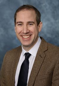
Jonathan Samet, MD
Division Head, Body Imaging Section Head, Musculoskeletal Imaging Department of Medical Imaging Ann & Robert H. Lurie Children's Hospital of Chicago Associate Professor of Radiology Northwestern University Feinberg School of Medici
Ann & Robert H. Lurie Children's Hospital of Chicago, Northwestern University Feinberg School of Medicine
© 2026 Medality. All Rights Reserved.
