Crohn’s Disease, Dr. Benjamin Spilseth, 02/03/22
HIDEInteractive Transcript
0:02
Hello and welcome to Noon Conferences hosted by MRI
0:04
online. Noon Conference was born out of the pandemic to
0:08
keep the radiology community connected with free live
0:10
conferences and make learning accessible from anywhere.
0:13
The overwhelmingly positive response to this has
0:15
only strengthened our mission to transform the
0:18
radiologist and thrive, learn, and thrive.
0:21
No matter the stage of your career or where in
0:23
the world you can practice. You can access the
0:25
recording of this conference and previous noon
0:27
conferences by creating a free MRI online account.
0:30
Link will be provided in the chat box.
0:32
Today, we are joined by
0:34
Dr. Benjamin Spilseth, one of our esteemed faculty
0:36
members who has also contributed essential mastery
0:39
series courses in gastrointestinal imaging, which
0:42
are available to MRI online premium members.
0:45
Both of which will be linked in the chat
0:46
box along with membership information.
0:49
Dr. Benjamin Spilseth is an Associate Professor of Radiology,
0:53
a Division Director of Abdominal Radiology at the University
0:56
of Minnesota, and Abdominal Imaging Fellowship Director.
1:00
He has published over 25 articles and book chapters
1:02
on abdominal and pelvic MRI, as well as being
1:05
a frequent presenter of lectures and workshops
1:07
at national societies including RSNA and SIR.
1:11
We truly appreciate him for joining us today
1:13
and being a part of our MRI online faculty.
1:16
A reminder that there will be a Q&A session at the end
1:19
of the lecture for any questions you may have for Dr. Spilseth.
1:23
Please use the Q&A chat feature to submit your questions.
1:25
We will get to as many as we can before our time is up.
1:28
With that being said, we welcome you.
1:30
1:30
Dr. Spilseth, please take it from here.
1:33
So, I'm Ben Spilseth.
1:34
I'm here at the University of Minnesota, and I'm
1:37
going to talk to you about Crohn's disease today.
1:42
So, first of all, this is an introductory talk.
1:46
It's going to be 40 minutes, and it's not enough
1:48
to get all the details, all the different
1:50
iterations you can see with Crohn's disease.
1:53
Um, as she just mentioned, I do have a more
1:57
fully detailed, um, case series
2:02
in the mastery series at MRI online.
2:05
If you want to check that out for more details, if you like
2:08
what you hear, or, um, you want some more experience working
2:11
with that, of course, um, that is a paid subscription piece.
2:14
So you'd have to pay for that, but, um,
2:17
but, uh, we'll, we'll get you kind of the,
2:19
the nuts and bolts of it with this talk.
2:21
So here, we're going to talk about the role of imaging,
2:24
some imaging techniques that we use for Crohn's disease.
2:27
And we'll also, um, talk about the key findings
2:32
and how to track severity, and then we'll do a
2:34
little bit of an intro to some of the advanced
2:36
topics that we talked about in the other series.
2:39
So we will get started here.
2:43
Um, so, you know, that's the
2:45
outline of what we're going to do.
2:46
Hopefully when we're all done, um, you, you're
2:48
not going to want to put me in that outline.
2:51
But if you do, there'll be a firing squad
2:53
of questions at the end of the series.
2:55
So you can go ahead and fire away at that time, or as we go
2:59
along, go ahead and enter your questions in the chat box and
3:02
we'll get to them as we go or towards the end of the time.
3:06
So Crohn's disease is
3:08
an idiopathic autoimmune disorder, and it
3:11
can involve any portion of the GI tract.
3:13
Um, it's typically a segmental, and, you know, as
3:16
you know, many cases have skipped lesions, so it can
3:20
involve anywhere from really the mouth to the anus.
3:23
Um, and oftentimes involves multiple regions.
3:27
Um, it's notably mostly in the terminal
3:29
ileum as, as kind of the classic location,
3:32
but any part of the bowel can be involved.
3:35
So why do we use, uh, radiology to image Crohn's?
3:39
You know, it's a good question because the
3:41
disease is often suspected or established with
3:43
direct visualization, colonoscopy, endoscopy,
3:46
um, can see Crohn's and make the diagnosis.
3:48
They can get biopsies, and the pathologist can oftentimes
3:52
diagnose the disease, but, and colonoscopy does remain
3:56
kind of the mainstay, uh, for disease monitoring.
3:59
But there are some reasons why we like imaging.
4:02
First of all, um, really getting the accurate diagnosis.
4:04
Sometimes you do need imaging to do that.
4:07
The big one is UC versus Crohn's.
4:09
On colonoscopy, there can be some overlap in how
4:12
they look and sometimes the pathologist can't
4:14
even tell on biopsy which one it's going to be.
4:18
Even experienced pathologists can have trouble
4:21
depending on what they're seeing on their slides.
4:23
So CT or MRI imaging is often needed
4:26
to exclude the small bowel involvement.
4:28
And that way, um, if it is small bowel, it
4:33
shouldn't be UC, it should be Crohn's disease.
4:35
This is also important because surgeons really hate
4:38
removing colons, um, in patients with a diagnosis
4:41
of UC when they really had Crohn's disease all
4:44
along because the outcomes are really poor.
4:46
If they do that, and they didn't know what they were getting
4:48
into, small bowel disease can be seen in over half of Crohn's
4:53
patients with a negative terminal ileum on ileocolonoscopy.
4:56
So sometimes they will do the colon and they'll see some
5:01
disease, but they think that the TI is not involved.
5:03
So they don't think there's small bowel disease.
5:04
Well, the truth is, they don't
5:06
know because they haven't seen it.
5:08
And so, um, doing MRI or CT is a great
5:11
way to view the rest of the small bowel.
5:15
Here's another reason why we do imaging
5:17
and that's monitoring the disease.
5:18
So after the disease has been established,
5:21
the gastroenterologists do need to track the severity
5:23
of inflammation and to monitor their treatment.
5:25
The symptoms that patients have don't really correlate
5:28
very well with what the actual disease activity is.
5:31
Um, they have to, you know, do counting
5:33
of stools and vague abdominal symptoms.
5:35
It can be.
5:36
It can be hard to really suss together how bad
5:39
their underlying disease is in these patients.
5:43
Capsule endoscopy is complimentary, but,
5:45
but CT and MRI are really getting widespread
5:47
use for monitoring Crohn's disease.
5:51
So we're going to start with, um,
5:53
talking about MRI and Crohn's disease.
5:55
And really it's all about technique.
5:58
And then it has to start with the
5:59
technique of how you do your exams.
6:00
If you don't get good MRIs, you can't give good
6:03
reports, you can't help the patients or your clinicians.
6:06
So we're going to spend a good
6:07
amount of time on the technique.
6:11
Part one of the technique is really the small bowel distention.
6:14
So when you do imaging, you have to give
6:18
the patient something to distend the bowel.
6:21
That way you can see the wall much better.
6:23
You can see if there's narrowing or strictures.
6:26
And if you don't get good distention,
6:27
you won't be able to see that.
6:29
So our protocol is we use three bottles
6:32
of, um, hyperosmolar agents.
6:34
So, Brexanolone, Volumin, there's other,
6:36
there's other ones out there.
6:38
And we usually do that over about 60 minutes.
6:40
Um, some people do it, uh, quicker than that.
6:43
And the key is that these agents have things
6:45
like sorbitol, mannitol, sucralose, things that
6:48
actually suck fluid into the gut as it goes through
6:51
because they're, um, hyperosmolar.
6:54
And so if you just give a patient water, you'll get good
6:57
distention of the stomach and duodenum and probably some of
6:59
the proximal jejunum, but by the time it makes it to the ileum,
7:03
your distention is gone and it's,
7:05
and it's reabsorbed into the body.
7:07
This is, this will go all the way through the ileum into
7:09
the colon, and that's why we use these agents.
7:13
You can also give a glass of water right before
7:14
getting on the table, just to, um, distend that,
7:17
that more proximal bowel, that can be helpful.
7:21
To get your distention.
7:22
So after we get distention, we have
7:24
another problem we have to deal with.
7:25
And that's the fact that the bowel
7:27
is a muscle that moves around.
7:29
And so you do need to give some sort of peristaltic
7:31
agent. In the U.S., that's typically glucagon.
7:34
And we do that for MRI. CT is fast enough
7:37
that you don't necessarily need that.
7:39
So we don't administer it for that.
7:41
So, um, at the University of Minnesota, we
7:44
do a scout film, and then we give IV glucagon
7:47
in, um, two divided doses of 0.5 milligrams to spread it out a little more.
7:50
So we give it right, you know, as we start and
7:52
then we give it again, um, before we get contrast.
7:55
A lot of places will just do a single dose.
7:58
Um, there's other routes you can do.
8:00
IM glucagon.
8:04
You can do, um, sublingual glucagon, although
8:06
the results don't typically look as well, and
8:08
I think some literature says that that's not
8:10
as, not as good as the IV or IM, uh, options.
8:14
Another thing we do for peristalsis, we do
8:16
try to image our patients in prone imaging.
8:18
And so, um, when you do prone imaging, what
8:23
happens is your bowel wall, um, your bowel doesn't
8:27
move as much if you're laying on your stomach.
8:29
And additionally, um, you get your images in coronal
8:35
images in fewer slices because the diameters decrease.
8:41
And so we try to image patients
8:42
in prone if they can tolerate.
8:44
Now if they can't tolerate it, we don't want to do that.
8:46
Um, because if motion is an issue and if
8:51
they're uncomfortable and they can't tolerate
8:52
the scan, it doesn't do anybody any good.
8:54
So only in the patients that are able to lay
8:55
prone, we do try to do prone imaging when possible.
8:59
Here's an example of the problems with bowel peristalsis.
9:02
If you look at this image, you see a lot of blurriness.
9:05
You can see there's bowel in here, but you
9:07
can't really get a lot of details from that.
9:09
And that's because this patient didn't, uh, get
9:11
the glucagon, their bowel was moving too much.
9:14
Um, this is a contrast-enhanced sequence.
9:17
When we look at the steady-state precession,
9:19
which isn't as motion sensitive because it's so
9:21
fast, we can see there's actually a big diseased loop
9:24
of bowel right here in the middle of the abdomen
9:26
that was really hard to see on, on this exam.
9:28
So, um, if we had a better, um, we can get that
9:33
image, but we can't get all the other sequences.
9:34
We really like to get the T2s, the T1s, pre and
9:37
post contrast, um, if you have bowel motion.
9:43
All right, the third thing we're going to talk
9:44
about is the sequences and this is also key.
9:47
There's a lot of guidance on which sequences
9:50
to do and you can look up the protocols and
9:51
literature in a bunch of different areas.
9:54
I'll tell you how we do it, though.
9:55
We do T2-weighted sequences.
9:57
With and without fat saturation.
9:58
And then we do, um, a steady-state free precession,
10:02
whether you have FISP or Fiesta, depending on your scanner.
10:05
Diffusion-weighted imaging, I do think is important.
10:07
And we do that.
10:08
And then we do pre- and post-contrast.
10:10
And optionally, more and more places are
10:13
doing some delayed imaging to help diagnose
10:15
fibrosis, and we'll talk about that.
10:17
And then you can do cine images
10:18
as well before you get glucagon.
10:21
Those, that produces some really nice pretty pictures.
10:23
Um, the diagnostic importance is probably not that
10:26
much, but our gastroenterologists do really like that.
10:28
So we do, we do cine images here as well.
10:32
So to help you look at those sequences, I'm going
10:34
to quickly go through a case and show you what
10:36
the sequences look like and how we use them.
10:39
And so here, starting at the top, we do some T2 images,
10:44
we do a HASTE image on our Siemens scanner, which
10:48
is kind of a faster half Fourier transform sequence.
10:52
And the, and you want to get
10:53
feed on these enterography exams because even with
10:56
the glucagon, if your sequence is too
10:59
slow, you're still gonna get motion artifacts.
11:02
Um, what you'll see here is you can
11:03
really see the bowel well, pretty well.
11:05
You can see that there's fluid
11:07
distending the lumen of this bowel.
11:08
So they did a good job with their preparation.
11:11
And you can also get the sense that there's some, a
11:13
loop of bowel wall that a loop of bowel that's sticking
11:16
down here in the location of the terminal ileum.
11:18
Here's your ileum and here's your
11:21
cecum.
11:23
So we're going to take a closer look at this
11:25
area and then talk about the other sequences.
11:26
One thing you'll notice is that this area of the
11:28
wall does look a little brighter than the skeletal
11:31
muscle, so it looks like it may be edematous,
11:33
but we want to do some things to confirm that.
11:35
And the way we typically confirm edema and look at it
11:38
is use the other T2 sequence, the fat-saturated T2.
11:41
And when we look at this, you can again see that that
11:44
bowel wall is indeed brighter than the skeletal muscle.
11:48
And when you have that, you know there's,
11:49
there's true edema and that's a marker of
11:51
inflammation, um, typical of Crohn's disease.
11:55
Now this isn't a specific, uh, appearance.
11:59
It doesn't have to be Crohn's.
12:00
Other types of ileitis can cause this, but
12:02
Crohn's disease, um, when you see it in that
12:04
location, it should be at the top of your mind.
12:08
Um, we also do T2 weighted images in the
12:10
axial plane to give us another look at things.
12:13
So after we do T2, um, we, we do the true FISP, which
12:18
is a type of steady state free precession image.
12:21
And one of the real advantages of the true
12:22
FISP is that it's not motion sensitive.
12:24
So if the bowel is moving a bit, you're
12:27
still going to get good images here.
12:28
Another big advantage is that you can really see the
12:31
lumen really well, so you notice there's no artifacts
12:34
in the lumen of the bowel here, so if there was a polyp
12:36
or some sort of intraluminal defect, you're going to
12:39
see it here on your T2 sequences because it's moving.
12:42
You get flow voids, kind of like the carotid, because you
12:46
can't, uh, you don't image things after you excite them.
12:48
They actually move out of plane.
12:49
So you get these flow voids in your bowel.
12:51
That's normal.
12:52
Don't call these polyps.
12:53
This is just, um, because of the,
12:55
the way the sequence is obtained.
12:57
But on TruFISP, you don't get those.
12:58
So that's why we do that series.
13:00
Then, um, we do the pre and post contrast.
13:02
So this is our key sequence and
13:06
it's a little more motion sensitive.
13:08
Um, but when it turns out well, you
13:10
get a really good look at things.
13:11
We like to do several time frames.
13:13
So we do a pre-contrast and then we do post
13:15
contrast, and we can see right away this bowel loop
13:18
is enhancing more than the adjacent bowel loops.
13:21
So that's a marker that there's some sort of
13:23
disease there, and it's inflamed in this case.
13:26
This has early enhancement, as you can see on this earlier
13:28
time series, and so that's a marker of an acute inflammation.
13:32
And when you're trying to detect early enhancement, it's a
13:34
key to look at it in relation to adjacent bowel loops.
13:39
So this is ileum, so we want to
13:40
compare it to other loops of ileum.
13:42
And what we see is that this is clearly brighter,
13:44
in addition to being thicker than the other loops of
13:47
ileum, and that's clearly an indicator of disease.
13:50
You don't want to compare ileum to jejunum,
13:52
because jejunum always looks like this.
13:54
This is normal jejunum, and you can see it has
13:56
normal wall thickness and normal thickening, but
13:59
it does enhance a little more than the ileum.
14:01
Don't, don't be confused by that.
14:02
That's a normal finding.
14:04
Although I see it called a lot of times and people that
14:06
aren't familiar with, with MRI (Magnetic Resonance Imaging) as disease.
14:09
Uh, but this is, this is normal.
14:11
If you want to confirm that it's normal, sometimes
14:13
you can look at your T2 series and see just how
14:16
normal looking this is in terms of nice thin
14:19
walls with the normal valvulae kind of entities.
14:23
Okay.
14:24
That's the detection piece on post-contrast.
14:27
Um, we do the multiple phases, including
14:30
the delayed phase, which helps with fibrosis.
14:32
If you have more enhancement later, it suggests fibrosis,
14:36
and we'll talk more about that later in the talk.
14:38
And then we do an axial phase to get another look.
14:40
In this case, the axial phase is helpful because
14:42
it does look like it's irregular and there's some
14:44
mucosal abnormalities on this patient,
14:48
um, and some ulceration indicating more severe disease.
14:52
Last sequence we'll talk about is the diffusion series.
14:54
And for diffusion, what I really focus on
14:57
is that the highest B value of the diffusion images that you get.
15:00
And what you're looking for there is an
15:02
area of increased signal in the bowel.
15:06
And in this case, that just indicates disease.
15:09
It doesn't necessarily indicate whether it's
15:11
acute or chronic, um, but it does indicate disease
15:14
and it can be used for troubleshooting at times.
15:17
It's also good to find some adjacent lymph nodes,
15:19
which pop out as being very bright on the future.
15:23
Okay, so that is that.
15:26
Now we'll go back to the PowerPoint quick.
15:36
All right, so back to our slides, um, a couple more
15:39
examples of what Crohn's disease looks like on MRI.
15:42
So this is a separate patient with ileal Crohn's.
15:45
You'll see enhancement.
15:46
This time it's just the inner wall that's enhancing.
15:49
We don't want to call that mucosal enhancement necessarily
15:51
because oftentimes the mucosa is actually sloughed off.
15:55
Um, so it's not truly always a mucosal enhancement, but
15:58
it is enhancing and that is an indicator of disease.
16:02
Additionally, this is which indicates some bowel disease.
16:06
And then, um, it's also edematous as well.
16:11
Here's a more mild case.
16:12
And in this case, this is your terminal ileum here.
16:15
And what you'll see is the, um, the wall is a
16:19
little thick and then maybe a little edematous, but
16:21
it can be hard to differentiate that from just a
16:23
little bit more prominent, normal terminal ileum.
16:25
So you need your other sequences
16:27
in this, in this, uh, scenario.
16:29
Uh, when we look at our diffusion series here.
16:32
What we see is that that is bright.
16:33
So this is where I find diffusion probably the most
16:36
helpful is differentiating these mild cases of disease.
16:39
Is that really a disease segment of bowel
16:41
or is it just a contraction problem?
16:44
And in this case, because it's so much more
16:45
enhancing than adjacent bowel, I think we can be
16:48
very confident that this is abnormal terminal ileum.
16:51
Similarly, it does show some increased hyper,
16:54
hyper-enhancement on the post-contrast images.
16:57
And so this is indeed a disease segment of bowel.
17:01
Okay.
17:03
Jejunal Crohn's obviously can also occur.
17:06
This is a case with multifocal areas of skip
17:09
lesions, so here you can see some thickening here
17:11
and some enhancement, and then this part in the left
17:14
lower quadrant is also thickened and enhancing.
17:17
When we look at the axial series on the same patient
17:20
you can see this jejunum is very thick and enhancing,
17:23
and there's some edema on the fat-saturated
17:26
series as well, indicating acute disease.
17:32
So that's the basics of MRI.
17:34
We'll move to the basics of CT.
17:36
So for preparation on CT, it's not as complex.
17:39
It's the same oral prep as the MRI.
17:42
And we don't do all the series and we don't
17:44
have to mess with glucagon because it's so fast.
17:46
We just get a single late arterial or enteric
17:50
phase image, and that's because that's the
17:52
time when the bowel is enhancing the most.
17:54
So you want to do this a little earlier than your classic
17:57
portal venous phase we do for most abdominal imaging.
18:01
Uh, the findings in CT are very
18:03
similar and analogous to MRI.
18:04
You just don't have as much to look at.
18:06
You just have the single sequence, and what you're
18:08
looking for here is primarily enhancing wall, wall
18:11
thickening, and then inflammation surrounding the wall,
18:14
as well as the complications you may see with Crohn's.
18:17
Here again, this colon is diffusely involved.
18:19
It's too thick, it's enhancing, it's irregular, and
18:22
there's even some fluid around the colon indicating
18:25
some substantial inflammation of the bowel.
18:30
So now we've gotten through diagnosing, and now
18:32
we're going to talk about monitoring disease.
18:33
And the question is, you know, why do we need imaging to
18:36
monitor this if we can do symptoms and colonoscopy?
18:40
Well, we've talked a little bit about the problems
18:42
with symptoms, but also, um, there's another factor
18:46
to consider. For one thing, the decision to
18:49
change drugs depends a lot on disease activity, and
18:53
the drugs they use these days are very expensive.
18:56
The immunomodulator infliximab and rituximab, etc.
19:01
They can cost tens to hundreds
19:02
of thousands of dollars a year.
19:04
And so the cost of, you know, $1,500 MRI compared
19:09
to that makes it really cost-effective to get to
19:12
use it to get a really good diagnosis and understand
19:14
what's going on with the patient's disease to pick the
19:17
right drug or decide if they even need drugs at all.
19:21
Additionally, we want to monitor disease for the detection
19:24
of complications such as fistulae and abscesses, which
19:26
aren't always apparent clinically or may not be suspected.
19:30
And then for surgical planning, monitoring
19:33
disease, understanding the full extent
19:35
of disease activity is really critical.
19:38
So as we do disease monitoring, one of the
19:40
questions we want to answer is, is there
19:42
inflammation that's active here or fibrosis?
19:45
The reason is inflammation is
19:46
treated differently than fibrosis.
19:48
Inflammation can be treated with immunologics,
19:51
infliximab, etc., or steroids, whereas
19:54
fibrosis is typically treated first with diet.
19:56
So if you have strictures, adjusting your diet
20:00
may help you get through that, or surgery if that
20:03
is, um, unable to be managed with diet, and immuno
20:09
modulators don't have a role once you become fibrotic
20:12
because you're no longer actively causing disease.
20:16
It's more than the sequela of disease
20:18
that's already happened, and surgery is
20:20
really the best bet to finally cure that.
20:23
So how do we tell inflammation versus fibrosis?
20:26
It's a challenge.
20:27
And the truth is, it's almost always a mixture of
20:30
both inflammation and fibrosis in Crohn's disease.
20:33
But we can find the predominant component, and we
20:36
can see that the degree of inflammation goes up
20:38
or down over time as we monitor these patients.
20:41
So how do we do that?
20:43
Inflammation, first of all, has higher T2 signal.
20:47
Um, and as we talked about before, if the signal of
20:50
the bowel is greater than the skeletal muscle on T2
20:53
series, that's an indicator that there is inflammation.
20:56
Inflammation also has ulceration and
20:58
blurred margins, especially when it's
21:00
more severe, and that's highly specific.
21:03
In the fibrosis side, the signal is dependent
21:05
on the amount of acute component on T2 series.
21:09
And the ulceration and blurred margins are
21:10
also not predictive of the amount of fibrosis
21:13
because those are markers of acute inflammation.
21:16
We can also look at the enhancement.
21:17
So early enhancement that doesn't
21:19
increase indicates inflammation.
21:21
Whereas if you have these progressive
21:23
enhancements, that indicates fibrosis.
21:25
And this is where we kick in those seven-minute images.
21:27
That's why we do that.
21:29
If you see more enhancement at seven minutes,
21:31
you're gonna, um, you're gonna think fibrosis is
21:34
a major component of the disease you're seeing.
21:37
So there's a good paper.
21:38
This is kind of a busy slide, but there's a good
21:40
paper showing that what I just said, that if
21:43
you have over 25 percent of enhancement gain at
21:46
seven minutes, you're going to see, you're going
21:48
to see patients do have significant fibrosis.
21:51
And depending on the T2 signal and that
21:54
enhancement gain, you can actually place
21:56
patients in this kind of two-by-two chart
21:59
in terms of whether they have fibrosis or inflammation.
22:02
And as we look at that in a little more detail,
22:04
what you see is what the T2 signal that's
22:07
going to tell you your inflammation component.
22:09
So if you have normal T2 signal, then
22:12
you have low inflammation and you're
22:14
going to be up on this part of the chart.
22:16
Whereas if you have bright T2 signal, you're
22:17
going to have more inflammation, higher
22:19
inflammation, and that'll put you down here.
22:21
And the next step is to look at the enhancements.
22:23
So if you have normal enhancement, you're going to
22:26
have lower fibrosis and only inflammation or no fibro
22:31
or low inflammation, whereas if you have delayed
22:33
enhancement and you have fibrosis on top of inflammation
22:37
on the bottom or without inflammation on the top.
22:39
So using T2 signal enhancement, you can really
22:42
decide how much inflammation and fibrosis you have.
22:46
So let's do one example here.
22:48
And there's many more that we went through
22:49
in the, um, the other talk I've done for MRI
22:52
online, but here's a case of mixed disease.
22:55
So if we look at this case, first thing
22:57
we're going to do is look at our T2 series.
22:59
And what we see here is the wall is thick.
23:01
So there's a disease segment, but it's also
23:03
a little bit brighter than skeletal muscle.
23:05
So that indicates there's some inflammation
23:07
when we look at it on the fat images.
23:10
We need that's important because fat can also cause
23:13
this bright signal on the T2 series, and on the
23:15
fat-saturated series we see it remains brighter
23:18
than skeletal muscle, so that's true inflammation.
23:20
So there's inflammation in this loop
23:21
of bowel, but what about fibrosis?
23:24
For that, we look to our contrast.
23:26
We look at the early enhancement here, and we see
23:28
it's enhancing a little bit, but the enhancement
23:31
increases as we get to that seven-minute delayed series.
23:35
And with that increased enhancement,
23:37
um, that indicates some fibrosis.
23:40
Now in the literature, they measure
23:41
that enhancement in practicality.
23:43
If you just look at it and you
23:44
see that it's clearly enhancing,
23:46
substantially more on the delayed series,
23:49
then you can say that there's also a fibrotic.
23:53
Um, beyond acute, uh, beyond the type of
23:56
inflammation, there's a severity of inflammation.
23:59
And there's been a lot of research that's
24:00
gone into developing severity scores.
24:03
There's these, the MARIA score and
24:05
Nancy score and others out there.
24:07
Um, the MARIA score has been validated, but
24:09
it's probably too complex for clinical use.
24:13
That being said, I'm going to just talk briefly about it.
24:15
And the MARIA calculation is very complicated.
24:18
They do a lot of noise adjustments and they
24:20
do a multi-factor weighted calculation.
24:24
And nobody in clinical practice really does this, but we can
24:28
get some key components from what the research has shown
24:31
about the MARIA score in terms of how severe inflammation
24:34
is, and this is how I use it: I look for the factors in
24:38
the MARIA score that were important, and I talk about those
24:41
in my reports, and I can relay that to my gastroenterologist.
24:44
So for one thing, if there's edema
24:45
or not, that tells you whether there's
24:47
severe, the severity of inflammation.
24:50
And this is just a plus-minus.
24:51
Is there, is there, is there not edema? More edema?
24:54
If there is edema, then that's indicated
24:56
there's some disease, at least.
24:58
Um, next thing I look at is ulceration.
25:00
So in this case, you see this
25:02
ulcerated component right here.
25:05
There's an irregularity in the wall.
25:07
Sometimes you'll see it on the T2 series.
25:09
Sometimes it'll be on the delayed series.
25:14
Or the contrast series, et cetera.
25:17
But whatever you see it best, if you see an
25:18
irregularity in the wall, you can call that ulceration.
25:21
And that indicates pretty severe
25:23
disease, severe inflammation.
25:25
That's the most, um, the most telling
25:28
factor of severe inflammatory disease.
25:32
The MARIA score also includes
25:33
wall thickness, which is helpful.
25:35
So if you're, you know, getting to five, six, um,
25:38
millimeters, you've got a really thickened wall.
25:40
Um, those are more severe disease cases.
25:43
And then you can, the early enhancement intensity
25:46
is also, um, is also a marker of inflammation.
25:50
And so if you have a lot of early enhancement,
25:54
that indicates more inflammation, and in the
25:57
MARIA score you measure that with our lives, etc.
26:00
And you use the brightest area, and in practice, you just
26:03
look at it and say if it's really bright and enhancing
26:06
in portions, it's probably more inflammation than if it wasn't.
26:10
And the last thing is the length. So MARIA doesn't directly
26:13
look at length, but we've shown independently
26:16
that length is a significant factor in
26:18
the degree of severity of Crohn's disease.
26:21
And so I try to report out the length of segments involved
26:24
to indicate severity and to monitor, but also to help the
26:28
gastroenterologist understand where the disease is in case
26:30
they can see it or in case surgery is needed to fix it.
26:35
Um, so that's severity.
26:36
Next, we're going to talk about the complications.
26:38
So, complicated disease is managed differently, so we want
26:42
to find things like fistulae, sinus tracts, and strictures.
26:45
And those can be hard to see or think about clinically,
26:48
but on imaging, we often are very helpful to the
26:51
clinicians in treating these patients that have them.
26:56
Uh, they can be found with CT or MRI,
26:59
and I'll talk a little bit about that.
27:02
So to understand these complications, you have to understand
27:05
that Crohn's disease is a transmural inflammatory disease.
27:09
And, um, because the whole bowel wall can get diseased,
27:12
then you can have strictures and fistulas and, and,
27:16
and problems from that so-called penetrating disease.
27:19
If you've broken through that bowel wall, they call
27:21
it penetrating disease, and now you're in a different
27:23
classification and a more severe type of Crohn's disease.
27:29
So there's, um, there's a kind of a
27:32
progression that Crohn's patients go through,
27:34
and I'm going to talk quickly about that.
27:36
So first of all, they can get strictures
27:38
because of the transmural inflammation.
27:40
It starts out as inflammation, and then over time it gets
27:45
either fibrotic or inflamed and narrows the lumen.
27:49
And once you've narrowed the lumen, eventually,
27:52
something's got to happen, and one of the most
27:54
common things is proximal dilation.
27:57
So as we're reading cases, when we see this narrow lumen
28:00
and proximal dilation, we can call that a stricture.
28:03
Be confident and slam dunk, say, you know, there's
28:05
a stricture here, especially on MRI where we
28:08
have multiple time points showing that narrowing.
28:11
If all we have is the narrowing and we don't have
28:13
the proximal dilation, we can't be as confident.
28:15
There's a, there's clearly a stricture because
28:17
it functionally may still be fine.
28:19
Um, and so this, we may have to hedge a little bit and say
28:22
that there's a possible stricture or something of that sort.
28:26
Once you've got, so here's an
28:27
example of an inflammatory stricture.
28:29
You can see, um, long segment. In this case, it could be
28:32
long or short, but this one's a longer segment, very
28:35
narrow lumen, and then proximal you've got some dilation.
28:38
You can see probably better here the degree that there is
28:40
clearly dilation proximal to that segment, and so this is
28:44
clearly a stricture, and that should be managed as such.
28:49
From strictures, you can develop fistulas, and it has to
28:52
do with not just the transmural inflammation but also
28:55
the fact that approximately that stricture that we get
28:58
pressure build-up, and that has to be released somewhere.
29:01
Ideally, it would go through the stricture, but sometimes
29:03
it goes through the weakened wall, and you get
29:07
fistulas or sinus tracts that develop or abscesses.
29:11
And so, this can be seen in the setting
29:13
of the proximal dilation or not.
29:15
Oftentimes, if you've developed the fistula, you no
29:17
longer have that proximal dilation because the, um,
29:21
the contents of the bowel have gone through,
29:25
they just haven't gone where you want them to.
29:28
So fistulas have a very characteristic appearance on Crohn's
29:31
patients, and that's because they're chronic, they're
29:34
chronic processes, and they often have a lot of scarring
29:37
and architectural distortion, and they basically stop
29:40
sucking all the bowel or other organs in the region towards it.
29:44
And so they have this kind of asterisk shape.
29:46
And if you see here, you can see this is a number of
29:50
segments of bowel that have been sucked together, and
29:52
centrally there's a little bit of anitis, and they're
29:54
all kind of tethered with this asterisk-shaped fistula.
29:57
This is a T2 series; you can also
29:59
see it on the post-contrast series.
30:02
Post-contrast here, T2, and they all kind of have this
30:04
nice little asterisk look to them, which is, I think,
30:07
very helpful in finding these fistulas, which often do
30:10
get mixed, I've noticed in private practice, um,
30:13
but people who just aren't used to seeing them.
30:18
So, um, finding strictures and fistulas brings
30:21
us to surgery, and, um, there are a couple
30:24
reasons why you might do surgery for Crohn's.
30:26
One is for the failure of medical management.
30:28
So, if there's no active inflammation to treat, but
30:31
there's fibrosis and the patient's still having symptoms,
30:33
sometimes surgery may be the only answer.
30:36
Or if the active inflammation is just no longer responding
30:39
to the medication, they've thrown the kitchen sink at it.
30:41
You can't get it better.
30:42
Sometimes they'll try to do surgery,
30:44
but there's also, um, if you have severe
30:47
stricturing or fistulae, those are other reasons
30:50
why the surgeon may need to be getting involved.
30:53
So when the surgeon does get involved, they often, the most
30:55
common thing is to do a hemicolectomy, taking the portion
30:58
of the right colon terminal ileum and then creating a new
31:02
terminal ileum or neo-terminal ileum with the terminal
31:05
ileum now connecting to the ascending colon somewhere.
31:10
And after we do that, we still end up
31:13
getting imaging to look for recurrence from
31:15
time to time because symptoms can recur.
31:17
It's a chronic condition that can relapse after surgery.
31:21
When they do have recurrence, it can occur anywhere, but
31:23
most commonly the anastomosis is seen, or if it's a patient
31:27
with an ostomy, that's another common site of recurrence.
31:31
It can be difficult to distinguish a mild post
31:34
operative inflammation, which isn't necessarily Crohn's
31:36
disease, from a true recurrence, and we have to look
31:39
at the severity to really make that distinction.
31:41
So here's an example of a recurrence.
31:43
This patient had a right hemicolectomy.
31:46
Here's the ascending colon, and here's the new terminal
31:50
ileum, and what you can see is that neo-terminal
31:52
ileum is a bit thickened, connecting up to there.
31:55
It's a bit enhancing.
31:56
And the degree of thickening and enhancing is,
31:59
is more than we'd expect postoperatively.
32:02
And so we would call that, uh, uh, some recurrent Crohn's
32:04
at that site, which can be verified on colonoscopy.
32:09
A couple more things before we're done here.
32:11
Um, one is to talk about the classic
32:13
question of is it Crohn's or is it UC?
32:16
So, um, there's some things that we've all learned in
32:18
medical school about this, and we'll go through that as
32:21
well as some other things that we can see on imaging.
32:23
So Crohn's does have the skip lesions, so it's not
32:25
confluent, whereas ulcerative colitis typically
32:28
classically starts at the anus and goes up
32:31
from there confluent. It may involve
32:35
portions of the colon or the entire
32:36
colon, and maybe the terminal ileum.
32:40
Crohn's can go from mouth to anus, whereas UC
32:43
is typically isolated to the colon, as we said.
32:46
Crohn's is transmural, which is why you get
32:48
fistulas and abscesses, whereas UC doesn't.
32:51
So, uh, you shouldn't expect to see the fistula and
32:54
abscess complications of UC in a typical patient.
32:58
Because it doesn't involve the whole wall,
32:59
it just involves more of the mucosal surface.
33:02
And that's why we see that.
33:05
Uh, also important to know that they both do
33:08
predispose to adenocarcinoma, so keep that in
33:10
your mind if you see masses forming or adenopathy
33:13
that's out of proportion to the inflammation.
33:16
And they both do have extraintestinal
33:17
manifestations, which we'll talk about next.
33:20
So Crohn's, especially colonic Crohn's,
33:23
and UC both have extraintestinal problems.
33:26
So, um, you can have musculoskeletal issues,
33:29
typically called, you know, seronegative spondyloarthropathy
33:32
that can involve the spine, can involve the SI
33:35
joints with an asymmetric inflammatory process.
33:38
And we see that in our MRN geography.
33:40
And so if you see over-enhancement of those
33:43
SI joints, don't forget that these patients
33:45
are at risk for that spondyloarthropathy and
33:48
we need to let the clinicians know about that.
33:51
PSC, primary sclerosing cholangitis, is another one.
33:55
It's typically seen with UC but can
33:56
also be seen in Crohn's disease.
33:58
And then along with that comes cholangiocarcinoma.
34:01
You can get stones and portal
34:03
vein thrombosis is another thing.
34:05
And especially in the acute stage,
34:07
keep your eye on that portal vein.
34:09
Make sure that you're looking at it to see
34:11
if there's thrombus because that can happen.
34:14
The skin findings are something we don't need
34:17
to worry about as radiologists, but can happen
34:19
and is important to know about, as well as some
34:21
ocular findings with episcleritis and scleritis.
34:25
PSC warrants a little bit more talk.
34:27
Um, so most PSC patients do have IBD.
34:31
So 75 percent of them have UC, 5 to 10 percent have Crohn's.
34:37
About 80, um, 15 to 20 percent that don't have IBD.
34:42
But that being said, PSC is uncommon in IBD patients.
34:45
So you can read a hundred, um, a hundred Crohn's patients,
34:49
and you'd only expect to see two patients that actually
34:51
have PSC. Nevertheless, we do want to look at it.
34:55
Cause that's a, that's still relative
34:57
to the population, a high rate of PSC.
34:59
So make sure you're looking in the liver,
35:01
looking for those peripheral areas of dilated
35:04
ducts or enhancement of the biliary tree.
35:07
If you see that, those could be early markers
35:09
of PSC, and that's very important for everyone
35:11
to know about before it gets too advanced.
35:15
So, uh, CT or MRI, um, what,
35:19
what can both detect on Crohn's?
35:21
Why would you choose one or the other?
35:23
Well, first of all, they both can detect inflammation.
35:25
They both are pretty good for fistulas and abscesses.
35:29
So I would be confident using both of them.
35:31
Strictures, you can see it on both, but I would
35:34
say MRI, because you get the multiple time points,
35:36
does have an advantage for evaluation of strictures
35:39
if that's what you know you're trying to solve.
35:42
But there's still reasons to choose
35:43
CT, so why would you choose that?
35:45
Well, it's much faster, it's available for emergencies,
35:48
whereas MRI can take a lot longer and it requires
35:51
a detailed protocol, and it's not available everywhere.
35:55
CT is more straightforward than MRI, and it's obviously
35:58
less expensive than MRI, although MRI, as I said
36:02
before, is cost-effective relative to no imaging.
36:07
Spatial resolution is probably a little
36:08
better on CT, but it's adequate on MRI.
36:11
And then you can have some error artifacts that can
36:13
cause problems on MRI that aren't an issue with CT.
36:17
What about MRI?
36:17
Why would you choose MRI?
36:19
Well, one big thing is there's no radiation, right?
36:22
Um, CT is high radiation, and that's especially
36:24
important for young Crohn's disease patients who may get
36:27
imaged, you know, half a dozen times a year for years.
36:30
Um, if you're getting radiation with each
36:32
one of those, that can affect you long term.
36:34
And so I really would like to see
36:37
MRI being used in those patients.
36:39
It gives you multiple time points on MRI,
36:41
which can be especially helpful for strictures.
36:45
You get really good soft tissue contrast, and you
36:48
can get more information from the T2 series,
36:51
the diffusion series, et cetera, which can help you
36:53
differentiate that acute and chronic inflammation.
36:56
So here's a nice case showing the
36:58
advantage of the multiple phases.
36:59
Any of you who've read CT have seen this before
37:02
when you have basically an intussusception
37:04
in the small bowel in the upper abdomen.
37:06
Sometimes it can be pretty, um, pretty significant
37:09
and you may need to talk about it in your report.
37:13
Well, in this case, you see a loop of bowel that's
37:16
intussuscepted into another loop of small bowel.
37:18
And you can see it on the post-contrast image
37:20
as well as this fat-saturated T2 series.
37:23
However, when we look at our regular T2
37:26
series done at a different time, it's not there.
37:29
And that indicates it's clearly a transient phenomenon.
37:32
It's of no clinical significance and probably
37:34
doesn't even need to be included in our report.
37:39
What about barium?
37:39
Barium used to be the mainstay of looking at Crohn's
37:42
disease, and small bowel follow-through can be helpful,
37:46
especially nowadays, if you're looking to characterize
37:50
or visualize fistulas, it can be used for that.
37:52
It's a dynamic imaging technique, so like MRI, but even
37:57
better, it can help you understand what's going on
38:01
with strictures in real time.
38:02
How severe are they and how functional are they?
38:04
Are they really restricting the flow of luminal materials?
38:09
Um, the problems are that it's just insensitive
38:11
for inflammation, especially mild inflammation,
38:14
and it's not as, as you, um, used currently.
38:18
So here's an example.
38:19
This is the same patient with a CT and a small
38:22
bowel follow-through. In the small bowel follow-through,
38:23
you can see there's a diseased segment of bowel here.
38:25
You can even see there's maybe a little
38:27
bit of erosion into the wall,
38:30
but you can't really tell how severe it is.
38:32
Whereas with CT, you get a lot more information,
38:34
and with MRI, you get even more still.
38:37
And so it's really gone to the wayside for the
38:40
most part now that MR enterography has taken hold.
38:44
So my recommendations on imaging are to
38:46
use CT or MRI for initial evaluation.
38:49
Either one, it really depends more
38:50
on the urgency and the setting.
38:52
If you're in the ER, and you're
38:54
questioning Crohn's, uh, I think CT is
38:56
totally reasonable, especially the first time.
38:59
However, when you're following it up, MRI
39:02
is typically recommended, especially for the younger
39:04
patients because of the radiation issues, but also
39:06
because you get a lot more information out of it.
39:09
If you're looking for strictures, start with MRI.
39:11
And consider barium if you need it.
39:13
And then for perianal disease, which is a whole
39:16
nother topic, MRI is really excellent for that.
39:19
And we actually have a whole nother course on perianal
39:21
disease, which we're not going to get into today, but it's
39:23
available, um, on the series on MRI online.
39:28
And then don't forget about barium
39:29
for troubleshooting from time to time.
39:31
It does come in handy.
39:33
So that's the basic stuff.
39:35
Um, we're not going to get too much into some of the
39:37
advanced topics we talk about in the full series.
39:40
Um, but in the advanced topics, we're talking more about,
39:43
you know, really using the up-to-date nomenclature.
39:46
Um, this is a great paper out of gastroenterology in
39:49
2018 from Dave Bruning at the Mayo Clinic, who's a
39:52
gastroenterologist along with a bunch of excellent
39:55
radiologists at the Society of Abdominal Radiology.
39:58
And, um, in this paper, they talk about
40:03
the types of patterns of enhancement and what the
40:05
indications or what the implications of that are
40:08
and how to identify those, as well as, you know,
40:11
the importance of wall thickening and what it means.
40:13
And, you know, we talked a bit about edema and
40:17
strictures, but it gives a little more detail
40:19
on how to handle, how to phrase things when you
40:22
have with or without upstream dilation, etc.
40:25
And then, um, characterization of the different
40:28
types of fistulas and what those things mean.
40:32
Um, so we get into that more in the other talk.
40:35
Additionally, we talked more about the Crohn's reporting.
40:37
So, you know, you want to include all the relevant
40:40
factors we talked about today, as well as ideally
40:43
some of the nomenclature that's in that other paper.
40:46
And this is best done if you can set up a
40:49
template that includes all the information,
40:51
and they do include one in that paper.
40:53
Everyone.
40:53
Which we walked through a little bit with some modifications
40:56
in that, in that other series, um, in which I, I try to
40:59
use that, that way of thinking when I dictate my cases.
41:04
So in conclusion, imaging is critical for
41:06
diagnosing and monitoring Crohn's disease.
41:09
CT is fast and accurate, but MRI has its
41:12
role and can give you more information.
41:15
And recognizing disease is the first step.
41:18
Monitoring is important, and detailed reports are key to
41:21
really help your gastroenterologist with monitoring disease.
41:26
So thanks for your attention, and we
41:27
will go to some questions at this point.
41:30
Great, thank you so much for sharing your
41:32
lecture with us today and for contributing your
41:34
Mastery Series courses that give radiologists a
41:36
shoulder-to-shoulder view of your reading skills.
41:38
At this time, we will open the floor
41:40
for any questions from our audience.
41:42
So we do have two in the Q&A, and then we have one
41:45
in the chat, so we'll begin with the one in the chat
41:47
just so we can get it out of the way.
41:50
So they were wondering if there are any hints to distinguish
41:53
early tumors, such as lymphoma or, oh, I don't know,
41:58
carcinoma versus severe inflammation on chronic disease.
42:01
Should we suggest potential malignancy in cases with high
42:03
degree of thickness and no changes despite treatment?
42:06
Yeah.
42:06
So, um, that can be a challenge.
42:09
Um, and the, um, the best thing is to
42:13
really understand what's happened over time.
42:15
And so oftentimes, ideally, you'll have those patients
42:18
that have been monitored over time, and if you get a one
42:21
shot, it can be a challenge, but if you've seen them over
42:23
time, um, you get a better sense, and then you correspond
42:27
with your, your, um, gastroenterologist and see what,
42:30
you know, what are they seeing, what have they seen,
42:32
because these patients have had colonoscopy or endoscopy.
42:36
And, um, if, so if it's in the colon or if it's in the
42:39
terminal ileum, they can typically look at it, um, and if
42:41
you're worried about it, you can, you can suggest that.
42:44
The real challenge is the, the mid small bowel stuff,
42:47
which is harder to get to, and you don't want to
42:49
go digging in there unless you absolutely have to.
42:53
So some things you can look for,
42:54
first of all, is the adenopathy.
42:56
You know, metastatic lymph nodes
42:58
have a different appearance.
42:59
They can be necrotic, they can look
43:00
irregular, they can be kind of ugly.
43:02
Uh, you do have reactive adenopathy
43:04
typically with Crohn's disease.
43:05
But when it starts to get really large or
43:07
really ugly looking, that's when I start to
43:10
talk about adenocarcinoma.
43:12
Um, the more nodular types of enhancement or, you
43:15
know, nodular, um, projections out of the wall,
43:18
uh, or into the lumen, those types of things can,
43:21
can be, um, more concerning for adenocarcinoma.
43:25
Uh, diffuse wall thickening.
43:27
I typically don't start thinking about it because you can
43:30
have really severe wall thickening with Crohn's disease.
43:33
And so, um, you need something more
43:35
than just wall thickening, um, to, to
43:38
to make that diagnosis.
43:39
Maybe if the wall thickening has gotten
43:41
worse over time, um, that sort of thing.
43:44
That's when you may think about it, despite it, like
43:47
they're on immunologics and most of it's improving,
43:49
but some area of wall thickening is getting worse.
43:51
That's a time you might think of it.
43:53
Um, lymphoma is a bit different, you know, for
43:56
lymphoma, I think it's a little easier to make
44:00
that differential, and the things you're looking for
44:02
there are more like the classic aneurysmal dilation.
44:06
So, if you have a really thick wall, but the
44:08
lumen isn't narrowed, then I'm starting to think
44:10
about small bowel lymphoma or large bowel lymphoma.
44:13
Often, usually, you'll have the kind of bulky
44:17
adenopathy as well. You know, true isolated bowel
44:20
lymphoma is pretty rare, and it's typically more
44:24
of that non-aneurysmal or aneurysmal dilation look.
44:27
And so, I find that helpful to make that
44:29
differentiation, but it can be a challenge for sure.
44:32
Okay.
44:32
We have another one.
44:33
Um, how to differentiate between edema and wall thickness?
44:38
So, they are really two different things.
44:39
Um, edema, you're looking at the, um,
44:44
for edema, you're looking at the signal.
44:47
So, edema is, how bright is it?
44:49
And in a case where you have
44:52
edema, what you're going to
44:55
do is compare it to the skeletal muscle.
44:57
And I'm just trying to go back
44:59
and find our edema slides here.
45:01
But, um,
45:04
here, you can see that this wall
45:06
is brighter than the muscle here.
45:10
And so, it's either fat or edema.
45:12
And so, when we look at our fat-saturated
45:14
series, if it's dark, then that means it's fat.
45:17
If it's bright, that means it's edema.
45:19
And so, that's how we make that distinction.
45:22
For wall thickness, we're just measuring the wall, and if
45:25
it's more than two or three millimeters, it's thickened.
45:27
And that doesn't necessarily indicate
45:30
an acute or chronic disease, but
45:33
it does indicate a diseased segment of bowel.
45:37
All right.
45:38
Uh, what's the best imaging feature to
45:40
define active disease?
45:43
Well, if you see ulceration, that's the best indicator.
45:47
Or if you see haziness
45:50
in the fat outside the bowel wall.
45:53
Sometimes, it looks blurry, especially
45:55
on your post-contrast images.
45:57
If you see one of those
45:59
two things, you can be very confident.
46:01
There's pretty extensive active disease.
46:03
Um, beyond that, you're looking for edema, and that
46:08
may indicate milder disease, but it does
46:11
suggest there's active disease if you have edema.
46:15
The enhancement of the wall itself is not as reliable
46:18
for indicating the activity of the inflammation.
46:22
So I try not to look at that as much as
46:24
those other factors that I just mentioned.
46:27
Thank you.
46:28
Why, in fibrosis, is the enhancement progressive?
46:32
So, throughout the body, when you have
46:34
scarring, you get progressive enhancement.
46:37
If you think about cholangiocarcinoma,
46:40
that's known to be more of a scarring-type malignancy.
46:43
And what you'll see is delayed enhancement
46:45
with cholangiocarcinoma.
46:48
It's the same with a desmoid-type tumor.
46:49
They enhance more later.
46:52
And it has to do with the fact that it's
46:54
not a hypervascular tumor or a hypervascular
46:57
process with a lot of edema and inflammation.
46:59
Instead, the contrast accumulates
47:02
over time in the areas of scar tissue.
47:05
And so, that's why we see it in Crohn's
47:07
disease, just like we see it elsewhere in the
47:09
body, as a delayed enhancement for fibrosis.
47:15
Thank you.
47:15
We have another one in the chat.
47:16
How do you approach enhancing bowel wall that
47:18
is not thickened and only mildly distended?
47:22
So, if you don't have a
47:26
diagnosis, there are two different situations.
47:28
One is if you know they have Crohn's, and the other
47:29
is if you're seeing it and don't know the cause.
47:32
It's an unknown case, basically,
47:34
and you're not sure what it is.
47:35
If that's the case and you just have
47:37
enhancement, it's very nonspecific.
47:40
It could be Crohn's, or any sort of
47:42
ileitis or inflammatory process, or infectious process.
47:46
But if you see it enhancing more
47:50
than adjacent bowel loops, then it is abnormal.
47:53
You need to call it some sort of
47:56
ileitis or colitis, but it's mild.
48:00
And the differential is much broader for that.
48:02
And so, you're not going to be able
48:05
to tell exactly what's going on necessarily.
48:07
You can just point to the fact that
48:08
there's some inflammation there.
48:11
All right, thank you so much.
48:13
We're going to leave it open for a few more seconds
48:15
before we give our final remarks.
48:20
We have another one.
48:21
It's, uh, Crohn's versus TB in the cecum and terminal ileum (TI).
48:26
Would you look at—oh goodness, I don't want to—
48:31
Yeah, so, um, Crohn's versus TB.
48:34
I mean, in Minnesota, that's not a very common differential.
48:38
And if you're in other parts of the
48:39
world, that certainly is more common.
48:42
So honestly, if you're in the cecum, the advantage
48:49
is that the next step is going to be a colonoscopy
48:53
if it's an unknown disease process. In the U.S.,
48:56
you're going to go to colonoscopy as the next step.
48:59
And they'll be able to tell a lot if it's Crohn's or,
49:03
or, um, TB, or some sort of malignancy in the cecum.
49:08
And so, really, our job in that scenario is really
49:11
just to get them to colonoscopy as the next step.
49:14
All right, thank you so much.
49:16
We're going to leave it open for
49:17
a few additional seconds.
49:20
There are some thank-yous and some praises
49:22
about your presentation in the chat.
49:25
Okay, we're going to leave it open for additional seconds.
49:30
All right, perfect.
49:34
So, as we bring our time together
49:35
to a close, I want to thank
49:36
Dr. Spilseth for this lecture, and thanks to all
49:38
for participating in our noon conference.
49:40
A reminder that you can access the recording of
49:42
today's conference and all other previous noon
49:44
conferences by creating a free MRI Online account.
49:48
If you'd like to access our Mastery Series courses,
49:50
case series, and much more educational content with
49:52
unlimited CME, you can sign up for a free seven-day
49:55
trial of our MRI Online Premium Membership.
49:58
You can learn more at mrionline.com,
50:00
at the URL in the chat box.
50:02
Be sure to join us next week on
50:04
Thursday, February 10th at 12 p.m.
50:05
Eastern time for a lecture on renal transplants
50:07
Doppler ultrasound evaluation.
50:10
You can register for that at MRI online.com
50:12
and follow us on social media at the MRI online
50:15
for updates and reminders on upcoming noon conferences.
50:18
Thanks again and have a lovely day.
50:20
Thanks.
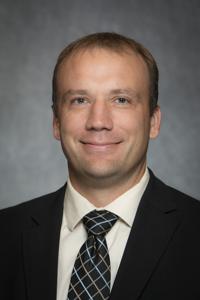
Benjamin Spilseth, MD, MBA, FSAR
Associate Professor of Radiology, Division Director of Abdominal Radiology
University of Minnesota
© 2026 Medality. All Rights Reserved.
