Training Collections
Library Memberships
On-demand course library with video lectures, expert case reviews, and more
Fellowship Certificate™ Programs
Practice-focused training programs designed to help you gain experience in a specific subspecialty area.
Ultimate Learning Pass
Unlock access to our full Course Library and all self-paced Fellowships.
Continuing Medical Education (State CME)
Complete all of your state CME requirements in one convenient place.
Noon Conference (Free)
Get access to free live lectures, every week, from top radiologists.
Case of the Week (Free)
Get a free weekly case delivered right to your inbox.
Case Crunch: Rapid Case Review (Free)
Register for free live board reviews.
Dr. Resnick's MSK Conference
Learn directly from the MSK Master himself.
Lower Extremities MRI Conference
Musculoskeletal Imaging
Emergency Imaging
Emergency GI & Abdominal Radiology FellowshipNEW!
PET Imaging
Pediatric Imaging
For Training Programs
Supplement your training program with case-based learning for residents, registrars, fellows, and more.
For Private Practices
Upskill in high growth, advanced imaging areas.
Compliance
NewTrack, fulfill, and report on all your radiologists' credentialing and licensing requirements.
Emergency Call Prep
Prepare trainees to be on call for the emergency department with this specialized training series.
Training Collections
Library Memberships
On-demand course library with video lectures, expert case reviews, and more
Fellowship Certificate™ Programs
Practice-focused training programs designed to help you gain experience in a specific subspecialty area.
Ultimate Learning Pass
Unlock access to our full Course Library and all self-paced Fellowships.
Continuing Medical Education (State CME)
Complete all of your state CME requirements in one convenient place.
Noon Conference (Free)
Get access to free live lectures, every week, from top radiologists.
Case of the Week (Free)
Get a free weekly case delivered right to your inbox.
Case Crunch: Rapid Case Review (Free)
Register for free live board reviews.
Dr. Resnick's MSK Conference
Learn directly from the MSK Master himself.
Lower Extremities MRI Conference
Musculoskeletal Imaging
Emergency Imaging
Emergency GI & Abdominal Radiology FellowshipNEW!
PET Imaging
Pediatric Imaging
For Training Programs
Supplement your training program with case-based learning for residents, registrars, fellows, and more.
For Private Practices
Upskill in high growth, advanced imaging areas.
Compliance
NewTrack, fulfill, and report on all your radiologists' credentialing and licensing requirements.
Emergency Call Prep
Prepare trainees to be on call for the emergency department with this specialized training series.
Lesson
Digital Breast Tomosynthesis Case Reviews
7 topics, 19 min. of video
Lesson
Tomosynthesis Guided Biopsy Case Reviews
3 topics, 15 min. of video
Lesson
DBT Biopsy of Calcifications Case Review
3 topics, 4 min. of video
0:00
Another screening mammogram. This was a case that was,
0:05
uh, recalled for a possible architectural distortion in the right breast.
0:09
So sometimes we'll we'll see on a, a full view,
0:13
an area that we question whether it's real, it's truly distorted or not.
0:16
We wanna have them come back to take an a closer look. So this was, um,
0:20
identified by the radiologist reading the screening mammogram.
0:26
I'm gonna show now the two D images. I'm gonna scroll through and see if we can
0:33
appreciate any distortion. I'm gonna circle,
0:36
this is where the finding was on her, on the two D image, but
0:41
when I'm scrolling through now on the three D imaging
0:45
and I'm following individual lines, I can scroll in and out of that area.
0:49
And there's not any single slice that I can pause on where I see a true
0:53
architectural distortion. So I think what, you know,
0:57
what we saw on that initial two D mammogram was, uh,
1:02
just the summation of normal overlapping tissue.
1:04
We did a Spock impression view also.
1:06
And similarly we see normal overlapping fibro glandular tissue.
1:11
There's no area that I can pause on that,
1:13
where I see a true architectural distortion. So this is another example of,
1:18
of how, um, of how uh, there are some,
1:22
there are many findings that with, with a tomosynthesis we can, uh,
1:27
clarify as being normal tissue and not pathology without requiring a biopsy.
1:35
We did an ultrasound at this area. Um, we showed normal,
1:38
normal breast tissue and the patient was, um, returned to screening.
Case 5: Possible Architectural Distortion
HIDEInteractive Transcript
0:00
Another screening mammogram. This was a case that was,
0:05
uh, recalled for a possible architectural distortion in the right breast.
0:09
So sometimes we'll we'll see on a, a full view,
0:13
an area that we question whether it's real, it's truly distorted or not.
0:16
We wanna have them come back to take an a closer look. So this was, um,
0:20
identified by the radiologist reading the screening mammogram.
0:26
I'm gonna show now the two D images. I'm gonna scroll through and see if we can
0:33
appreciate any distortion. I'm gonna circle,
0:36
this is where the finding was on her, on the two D image, but
0:41
when I'm scrolling through now on the three D imaging
0:45
and I'm following individual lines, I can scroll in and out of that area.
0:49
And there's not any single slice that I can pause on where I see a true
0:53
architectural distortion. So I think what, you know,
0:57
what we saw on that initial two D mammogram was, uh,
1:02
just the summation of normal overlapping tissue.
1:04
We did a Spock impression view also.
1:06
And similarly we see normal overlapping fibro glandular tissue.
1:11
There's no area that I can pause on that,
1:13
where I see a true architectural distortion. So this is another example of,
1:18
of how, um, of how uh, there are some,
1:22
there are many findings that with, with a tomosynthesis we can, uh,
1:27
clarify as being normal tissue and not pathology without requiring a biopsy.
1:35
We did an ultrasound at this area. Um, we showed normal,
1:38
normal breast tissue and the patient was, um, returned to screening.
Report
Faculty
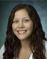
Emily B. Ambinder, MD
Assistant Professor - Breast Imaging Division
The Russell H. Morgan Department of Radiology and Radiological Science, Johns Hopkins Medicine
Tags
Ultrasound
Tomosynthesis
Neoplastic
Mammography
Breast
© 2026 Medality. All Rights Reserved.
