Training Collections
Library Memberships
On-demand course library with video lectures, expert case reviews, and more
Fellowship Certificate™ Programs
Practice-focused training programs designed to help you gain experience in a specific subspecialty area.
Ultimate Learning Pass
Unlock access to our full Course Library and all self-paced Fellowships.
Continuing Medical Education (State CME)
Complete all of your state CME requirements in one convenient place.
Noon Conference (Free)
Get access to free live lectures, every week, from top radiologists.
Case of the Week (Free)
Get a free weekly case delivered right to your inbox.
Case Crunch: Rapid Case Review (Free)
Register for free live board reviews.
Dr. Resnick's MSK Conference
Learn directly from the MSK Master himself.
Lower Extremities MRI Conference
Musculoskeletal Imaging
For Training Programs
Supplement your training program with case-based learning for residents, registrars, fellows, and more.
For Private Practices
Upskill in high growth, advanced imaging areas.
Compliance
NewTrack, fulfill, and report on all your radiologists' credentialing and licensing requirements.
Emergency Call Prep
Prepare trainees to be on call for the emergency department with this specialized training series.
Training Collections
Library Memberships
On-demand course library with video lectures, expert case reviews, and more
Fellowship Certificate™ Programs
Practice-focused training programs designed to help you gain experience in a specific subspecialty area.
Ultimate Learning Pass
Unlock access to our full Course Library and all self-paced Fellowships.
Continuing Medical Education (State CME)
Complete all of your state CME requirements in one convenient place.
Noon Conference (Free)
Get access to free live lectures, every week, from top radiologists.
Case of the Week (Free)
Get a free weekly case delivered right to your inbox.
Case Crunch: Rapid Case Review (Free)
Register for free live board reviews.
Dr. Resnick's MSK Conference
Learn directly from the MSK Master himself.
Lower Extremities MRI Conference
Musculoskeletal Imaging
For Training Programs
Supplement your training program with case-based learning for residents, registrars, fellows, and more.
For Private Practices
Upskill in high growth, advanced imaging areas.
Compliance
NewTrack, fulfill, and report on all your radiologists' credentialing and licensing requirements.
Emergency Call Prep
Prepare trainees to be on call for the emergency department with this specialized training series.
5 topics, 23 min.
3 topics, 14 min.
2 topics, 7 min.
2 topics, 2 min.
5 topics, 21 min.
Introduction to CEM Case Review
4 m.How to Use the Case Viewer to Best Evaluate the Images in this Course
2 m.Minimal Background Parenchymal Enhancement and Artifacts
5 m.Moderate/Marked Background Parenchymal Enhancement
9 m.Dense Breasts, Mild Background Parenchymal Enhancement, Axillary Enhancement Artifact
3 m.4 topics, 15 min.
13 topics, 57 min.
Rim Enhancement of Hematoma Obscuring Enhancing Cancer
6 m.High Conspicuity Enhancing Mass of Invasive Lobular Carcinoma
6 m.Low Conspicuity, Delayed Enhancement of Mucinous Carcinoma
6 m.Rim Enhancing Invasive Ductal Carcinoma in Patient With Implants
4 m.Low Conspicuity Invasive Ductal Carcinoma in Extremely Dense Breasts
4 m.Multiple Masses and Non Mass Enhancement of Invasive Lobular Carcinoma
6 m.Focal Non-mass Enhancement of Invasive Ductal Carcinoma
6 m.Linear Non-mass Enhancement of Ductal Carcinoma In Situ
3 m.Non-mass Enhancement of Ductal Carcinoma In Situ; Using CEM For Biopsy Planning
7 m.Calcified Ductal Carcinoma In Situ With No Enhancement
3 m.Invasive Ductal Carcinoma as Linear Non-mass Enhancement
4 m.Limitations of Background Parenchymal Enhancement in Cancer Staging
4 m.True Positive CEM in Post-Surgical Breast
5 m.1 topic, 1 min.
0:00
This is a case of an 81-year-old woman,
0:03
uh, who presented to our institution
0:05
for left breast cancer staging.
0:07
It was an invasive ductal cancer, grade
0:09
two, ER/P positive, HER2 equivocal.
0:14
These are her all low energy images, fatty or
0:17
scattered triradiate glandular densities on the left, uh,
0:21
focal asymmetry, maybe a little mass with the clip.
0:25
On the right.
0:26
It's hard to see here, but, uh, you will
0:29
have a benefit of magnifying that area.
0:32
There are.
0:32
There is an area of regional micro
0:34
calcifications, so here they are.
0:38
However, looking at the, uh, contrast-
0:41
enhanced mammography at the combined images
0:44
of the contrast-enhanced mammography,
0:46
we don't see any asymmetry at all on the right.
0:49
So in the area of those calcifications,
0:51
there is no enhancement at all.
0:53
Although we do see low to moderate conspicuity
0:56
mass on the left, and that's the known cancer.
1:00
When we do an ultrasound.
1:02
So we see it as expected in the
1:03
regular mass with the clip in place.
1:06
So this is an MRI—breast MRI—and
1:09
she happened to have an MRI also.
1:11
And on the right we see the left
1:14
enhancing cancer on the, uh, maximal
1:18
intensity projection image on the left.
1:20
So this is her enhancing cancer.
1:24
On the right we see, uh, one
1:27
duct with the T1 hyperintense material in it.
1:31
So it's either blood or it's, uh, protein-
1:34
tenaceous material, but it doesn't really
1:36
enhance because this is actually a MIP of a post-
1:39
contrast image, and this is a T1 shine through.
1:42
You see that on pre-contrast image, it's also bright.
1:46
Apart from that enhancing duct, the MRI
1:49
didn't really show us any pathology either.
1:51
So the point of me bringing up
1:53
the MRI here is that neither
1:56
CEM nor MRI showed any focal pathology
2:00
in the area of concern—in the area of
2:02
those mammographic microcalcifications.
2:05
However, as Dr. Phillips pointed out, CEM is a combined
2:10
exam, and the suspicious findings on low energy images
2:15
are enough to do a biopsy and to do further workup,
2:19
even if there is no enhancement on recombined images.
2:25
So we did do a stereotactic-guided biopsy
2:28
of the right, uh, microcalcifications,
2:30
and it was DCIS, intermediate grade.
2:34
On surgical pathology, she had a segmental
2:36
mastectomy, which confirmed grade 2
2:38
DCIS of 5 millimeter in diameter.
2:41
So it actually was not as big
2:43
as the area of calcifications.
2:45
But the point is, uh, that especially with
2:47
the calcifications, if the calcifications
2:50
are suspicious enough, even if they don't
2:52
enhance, they have to be biopsied.
Interactive Transcript
0:00
This is a case of an 81-year-old woman,
0:03
uh, who presented to our institution
0:05
for left breast cancer staging.
0:07
It was an invasive ductal cancer, grade
0:09
two, ER/P positive, HER2 equivocal.
0:14
These are her all low energy images, fatty or
0:17
scattered triradiate glandular densities on the left, uh,
0:21
focal asymmetry, maybe a little mass with the clip.
0:25
On the right.
0:26
It's hard to see here, but, uh, you will
0:29
have a benefit of magnifying that area.
0:32
There are.
0:32
There is an area of regional micro
0:34
calcifications, so here they are.
0:38
However, looking at the, uh, contrast-
0:41
enhanced mammography at the combined images
0:44
of the contrast-enhanced mammography,
0:46
we don't see any asymmetry at all on the right.
0:49
So in the area of those calcifications,
0:51
there is no enhancement at all.
0:53
Although we do see low to moderate conspicuity
0:56
mass on the left, and that's the known cancer.
1:00
When we do an ultrasound.
1:02
So we see it as expected in the
1:03
regular mass with the clip in place.
1:06
So this is an MRI—breast MRI—and
1:09
she happened to have an MRI also.
1:11
And on the right we see the left
1:14
enhancing cancer on the, uh, maximal
1:18
intensity projection image on the left.
1:20
So this is her enhancing cancer.
1:24
On the right we see, uh, one
1:27
duct with the T1 hyperintense material in it.
1:31
So it's either blood or it's, uh, protein-
1:34
tenaceous material, but it doesn't really
1:36
enhance because this is actually a MIP of a post-
1:39
contrast image, and this is a T1 shine through.
1:42
You see that on pre-contrast image, it's also bright.
1:46
Apart from that enhancing duct, the MRI
1:49
didn't really show us any pathology either.
1:51
So the point of me bringing up
1:53
the MRI here is that neither
1:56
CEM nor MRI showed any focal pathology
2:00
in the area of concern—in the area of
2:02
those mammographic microcalcifications.
2:05
However, as Dr. Phillips pointed out, CEM is a combined
2:10
exam, and the suspicious findings on low energy images
2:15
are enough to do a biopsy and to do further workup,
2:19
even if there is no enhancement on recombined images.
2:25
So we did do a stereotactic-guided biopsy
2:28
of the right, uh, microcalcifications,
2:30
and it was DCIS, intermediate grade.
2:34
On surgical pathology, she had a segmental
2:36
mastectomy, which confirmed grade 2
2:38
DCIS of 5 millimeter in diameter.
2:41
So it actually was not as big
2:43
as the area of calcifications.
2:45
But the point is, uh, that especially with
2:47
the calcifications, if the calcifications
2:50
are suspicious enough, even if they don't
2:52
enhance, they have to be biopsied.
Report
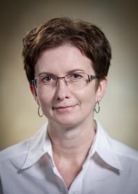
Faculty
Olena Weaver, MD
Associate Professor
Department of Breast Imaging, The University of Texas MD Anderson Cancer Center
Tags
Oncologic Imaging
Neoplastic
Mammography
Diagnosis & Staging
Breast
© 2026 Medality. All Rights Reserved.