Training Collections
Library Memberships
On-demand course library with video lectures, expert case reviews, and more
Fellowship Certificate™ Programs
Practice-focused training programs designed to help you gain experience in a specific subspecialty area.
Ultimate Learning Pass
Unlock access to our full Course Library and all self-paced Fellowships.
Continuing Medical Education (State CME)
Complete all of your state CME requirements in one convenient place.
Noon Conference (Free)
Get access to free live lectures, every week, from top radiologists.
Case of the Week (Free)
Get a free weekly case delivered right to your inbox.
Case Crunch: Rapid Case Review (Free)
Register for free live board reviews.
Dr. Resnick's MSK Conference
Learn directly from the MSK Master himself.
Lower Extremities MRI Conference
Musculoskeletal Imaging
PET Imaging
Pediatric Imaging
For Training Programs
Supplement your training program with case-based learning for residents, registrars, fellows, and more.
For Private Practices
Upskill in high growth, advanced imaging areas.
Compliance
NewTrack, fulfill, and report on all your radiologists' credentialing and licensing requirements.
Emergency Call Prep
Prepare trainees to be on call for the emergency department with this specialized training series.
Training Collections
Library Memberships
On-demand course library with video lectures, expert case reviews, and more
Fellowship Certificate™ Programs
Practice-focused training programs designed to help you gain experience in a specific subspecialty area.
Ultimate Learning Pass
Unlock access to our full Course Library and all self-paced Fellowships.
Continuing Medical Education (State CME)
Complete all of your state CME requirements in one convenient place.
Noon Conference (Free)
Get access to free live lectures, every week, from top radiologists.
Case of the Week (Free)
Get a free weekly case delivered right to your inbox.
Case Crunch: Rapid Case Review (Free)
Register for free live board reviews.
Dr. Resnick's MSK Conference
Learn directly from the MSK Master himself.
Lower Extremities MRI Conference
Musculoskeletal Imaging
PET Imaging
Pediatric Imaging
For Training Programs
Supplement your training program with case-based learning for residents, registrars, fellows, and more.
For Private Practices
Upskill in high growth, advanced imaging areas.
Compliance
NewTrack, fulfill, and report on all your radiologists' credentialing and licensing requirements.
Emergency Call Prep
Prepare trainees to be on call for the emergency department with this specialized training series.
Lesson
Pre-Course Activities
1 topic, 6 min.
Lesson
TAVR Indications, Protocols and Devices
6 topics, 26 min.
Lesson
Standard Aortic Root Measurements for TAVR
10 topics, 47 min.
Measuring the Annulus
6 m.Annular Measurement, Manual Method Case Review
8 m.Annular Measurement, Software Assisted Case Review
4 m.Additional Measurements of the Aortic Root
4 m.Coronary Artery Heights Case Review
6 m.Sinus of Valsalva Widths and Heights Case Review
9 m.Fluoroscopic Planning Case Review
3 m.Valve-in-Valve
6 m.Valve-in-Valve Post TAVR Case Review
2 m.Valve-in-Valve Case Review
4 m.Lesson
Ancillary Pre-TAVR Aortic Root Findings
11 topics, 33 min.
Aortic Valve Calcium Scoring
3 m.Aortic Valve Calcium Scoring Case Review
4 m.Annular Calcifiations
3 m.Annular Calcification Case Review
3 m.Bicuspid Aortic Valve
6 m.Bicuspid Aortic Valve (2 Sinus) Case Review
2 m.Bicuspid Aortic Valve (3 Sinus with Fusion) Case Review
3 m.Bicuspid Aortic Valve (2 Sinus) Post TAVR Case Review
3 m.Coronary Disease
3 m.Membranous Septal Length
4 m.Membranous Septal Length Case Review
4 m.Lesson
Vascular Access Planning for TAVR
3 topics, 17 min.
Lesson
TAVR Complications
5 topics, 21 min.
TAVR Complications
HIDEInteractive Transcript
Report
Faculty
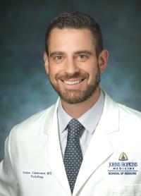
Stefan Loy Zimmerman, MD
Associate Professor of Radiology and Radiological Science
Johns Hopkins Medicine Department of Radiology and Radiological Science
Tags
Vascular Imaging
Vascular
Idiopathic
Congenital
Cardiac valves
Cardiac
CTA
CT
Acquired/Developmental
© 2026 Medality. All Rights Reserved.
