Spine Degeneration & Inflammation, Dr. Marcelo de Abreu (1-19-23)
HIDEInteractive Transcript
0:01
Hello and welcome to New conference hosted
0:04
by MRI online. We create a
0:07
new conference when the pandemic hit as a way to connect
0:10
the global Radiology community Through free live educational
0:13
conferences that are accessible for
0:16
all it's become an amazing weekly opportunity to
0:19
learn alongside Radiologists from all around the world and we
0:22
encourage you to ask questions and share ideas to
0:25
help the community Learn and Grow.
0:27
You can access the recording of today's conference and
0:30
previous new conferences by creating a free
0:33
MRI online account. The link will be provided in
0:36
the chat box.
0:38
You can also sign up for a free trial of our premium membership to
0:41
get access to hundreds of case-based micro learning
0:44
courses across all key Radiology subspecialties. Learn
0:47
more at mrnline.com.
0:51
Today we are honored to welcome Dr. Marcello day
0:54
Abreu.
0:55
for a lecture on spine degeneration and inflammation
0:59
Dr. Abreu is a member of international skeletal radiology
1:02
and staff at Davos idkd annual
1:05
course. He completed his msk fellowship
1:08
and neuroradiology fellowship at UC, San
1:11
Diego.
1:12
He has now head of radiology at Hospital May Day
1:15
do.
1:16
At the end of the lecture, please join Dr. Abreu in
1:19
a Q&A session where he will address questions you
1:22
may have on today's topic. Please remember to use
1:25
the Q&A feature to submit your questions so we can get
1:28
to as many as possible before time is up.
1:31
And with that we're ready to begin today's lecture Dr. Abreu.
1:34
Please take it from here.
1:36
Okay guys, so that's talk about
1:39
these generation and inflammation.
1:43
fine
1:46
So this is a very for me
1:49
very interesting topic.
1:51
We see this every day a lot
1:54
of cases.
1:56
Of spine generation and information. I'm
1:59
beginning the presentation. They're showing you.
2:02
example of two individuals
2:06
with no symptoms and
2:11
some definitive changes and I
2:14
will ask you to guess the age
2:17
of both.
2:24
So both individuals here. They have 75
2:27
year old, both are male.
2:31
and we see a lot of
2:34
a lot of difference between the definitive process
2:39
in the same in the individuals with same
2:42
age and that's because
2:45
it varies a lot the difference of
2:48
process.
2:49
and
2:52
it has to do with genetic component.
2:55
Yeah, and that's very specific for each individual.
2:58
The genetic component can be at the
3:01
molecular level talking about
3:04
collagen type.
3:07
Or an atomic level for example, if the
3:10
individual have a use it
3:13
segment for transitional fat.
3:17
And also the definitive process could be
3:20
caused by overuse for micro trauma.
3:24
So that's why we have
3:27
a lot of variation.
3:29
between individuals
3:34
about the anatomy mainly the weight
3:37
that
3:40
will reach the spine will
3:44
Reach the interval people disk 70%
3:47
of the weight.
3:50
So most of these native process
3:53
they begin in the spine at the
3:56
disk level in after that.
4:00
At the facet joints usually the facet trying
4:03
to process.
4:05
I
4:07
the secondary to a problem in the
4:12
Interior portion of the spine. Okay so
4:15
that we need to know that to understand all
4:18
the biomechanics and how
4:21
the things will develop in spine. Okay.
4:24
We have something called the degenerative Cascade.
4:28
And that begins with the
4:31
he died dehydration.
4:34
Of the disc and that's has to
4:37
do with the end played
4:40
that nourish the disc. It's a
4:43
collage. It's a cartilage layer
4:46
and near the court football
4:49
that we cannot usually see and that
4:52
will
4:53
give a nutrition for
4:56
the disk.
4:57
and a problem at that
5:00
include will
5:02
Begin, the definitive Cascade. Yeah that
5:05
usually begins.
5:07
yeah, the third deck of Life
5:10
MRI can show
5:14
that beginning with dehydration of
5:17
internal portion of the local posters here
5:20
that we can see
5:22
and then
5:24
after the dehydration will have
5:27
features.
5:29
And the structuring of this so called
5:32
alteration osteophyte in some
5:35
cases will have instability. So this will
5:38
start at the third decade of Life can start
5:41
before if the patient has like spondylolises
5:44
has a
5:47
trial or Reason young athlete
5:50
team athlete could
5:53
start in the in the second decade
5:56
of life. Okay?
6:00
I'll show you an example of that.
6:04
We can see here dehydration of
6:07
Calgary or 4 and 405.
6:12
This could be normal with 47 year old.
6:15
But the patient had a presented with
6:18
left Randy left radical property.
6:21
Okay, and then with this coronal sequence
6:24
that is the food sensitive sequence. We
6:27
can see animals fibrosis
6:30
feature here a high signal
6:33
intensity.
6:35
and
6:37
because of the the sensitivity to inflammation and
6:40
edema, we can see edema outside
6:44
that this year so
6:47
When you have a chronic.
6:50
And it was fibrosis there here in
6:53
the below level. But this one
6:56
in our Field Four is a cute we
6:59
can see that in a zoom image here because of
7:02
the information around that.
7:06
And using a diffusion sequence
7:09
that we are using right now a
7:12
lot with the sometimes in
7:15
routine Mr. But sometimes with the exam
7:18
the pair of calling neurography we can
7:21
actually see the nerve itself. Okay. So
7:24
for example, this is an axio.
7:28
Diffusion we put psif sequence
7:31
showing the inflamic nerve
7:34
the left LT root.
7:39
And just adjacent to the animals fiber
7:43
there. This is a movie.
7:46
showing
7:48
showing that
7:50
Okay, the relationship between information
7:53
and radicalopathy.
7:57
In the scenario of digital to
8:00
this Cascade, okay.
8:03
So it's important to look that when we
8:06
look at this active spines.
8:08
There are paper showing that.
8:13
this
8:16
when we inject contrast we can have that Vision
8:19
we can see the information process
8:22
in addition to this.
8:25
With acute analysts of
8:28
fiber pair. Okay, there are
8:31
usually three types of pairs the ones
8:34
that cause most inflammation are the consensic
8:37
tears.
8:39
The radial tear usually does not
8:42
cause inflammation the radio care.
8:45
Allows communication of the nuclear proposals with
8:48
the animals and is a kind of free herniated
8:51
disc.
8:53
and
8:55
okay.
8:57
so this is
9:00
a thing that when we are looking at that that exam.
9:05
We see a lot of tradition and we
9:08
should.
9:09
Try to find the point of information that
9:12
can come.
9:15
inside the disc subchondragon
9:18
Facet Joint into spinous process
9:21
joint so there are many points that
9:24
we can see the compensation of
9:27
additional tips spine. Okay, and
9:30
we need to set our protocols
9:33
to find that.
9:35
and that's why we are using lot the
9:39
the food sensitive sequence. We are using steer
9:42
or T2 headset in this.
9:45
sagittal plane and also in the coronal plane
9:49
To look to those hiding areas of
9:52
information.
9:54
This is an example here.
9:57
Yeah, patient seven years old enough pain.
10:00
We have a transitional vertebra. So
10:03
we have a patient with a
10:06
congenital defect the congenital level
10:09
defect so there is
10:12
one transitional value with
10:15
Articulation with the sacrum here
10:18
some kind of degenerative process and we
10:21
use the fluid sensitive sequence to see
10:24
that inflammation. Okay.
10:29
And we can get we can catch that see a lot
10:32
of Edema here showing that beside the
10:36
the OA there is a
10:39
lot of bone marrow edema here and probably
10:42
related to symptoms. Okay,
10:45
so it's very important to use those people.
10:49
Also in the same patient see we can
10:52
find.
10:54
the modic type 1 change at the right and a
10:59
Inflammatory process around the
11:02
discount trio4 on the
11:05
right. So we probably have here and
11:08
our analyst here with inflammation and
11:11
that could be related to a the
11:14
right equal opportunity. So
11:17
we pay attention on those sequences.
11:21
Okay.
11:24
Also, it's good to remind when we have that transition of
11:27
bad people. So we have an atomic
11:30
effect.
11:31
Will alter all the biomechanics okay
11:34
of the spine, so we have an increase
11:37
it.
11:39
Angulation between L4 and
11:42
L5 in this patient with fuse it transitional.
11:45
We have the bread on the right and that will
11:48
create dispose to
11:52
listis dictionary process can also
11:56
for I mean
11:58
of the canal, okay.
12:01
So it's important to understand.
12:04
The biomechanics when you look at this
12:07
time.
12:08
so the digital Cascade
12:11
and we can
12:13
have some micro instability. Okay,
12:16
and in the beginning
12:19
of the process when we have a black disc.
12:23
and just after the it dehydration
12:26
completes we this black
12:29
this
12:31
They are soft they are usually soft
12:34
and they develop some Michael disability that
12:37
usually we don't catch that with.
12:41
Conventional MRI we need to do a dynamic exam.
12:45
and here is an example of a dynamic MRI that
12:48
we
12:49
have a protocol use when with the open board
12:52
okay to do that.
12:55
and then we can see the
12:59
the instability on the on the right here
13:02
when we apply flexion to the exam. So
13:05
sometimes we have a boat in
13:08
this the black this and we have instability, but
13:11
we don't see it. And then with the
13:14
dining camera I can in this example showing that
13:17
them this is exist. We
13:21
are adding this new neurography protocol.
13:24
Okay for the
13:27
For the routine MRI, we
13:30
use a 3 minute sequence
13:33
a coronal steer space. Okay. It's a
13:36
fast sequence and it's a good gives you
13:40
a good clue.
13:42
About the nerve Roots. So you have
13:45
a definitive spine. You want to know the level of
13:48
the problem and then
13:50
You can have a group with this sequence here
13:53
see the roots.
13:56
and the ganglion the sciatic nerve
13:59
bilateral
14:01
and when we have on
14:04
your native use if you want to to look at
14:07
and
14:09
And do a complete protocol this is
14:12
the complete neurology protocol this sequence here,
14:15
and I'm sure is the psif but
14:18
the diffusion cpacy showing the relationship
14:21
between the native. Yes that we
14:24
can see on the right health for a fight and the
14:27
relationship with L4 and L5 group.
14:30
So you can tell what route
14:33
in this case the out the right L4 route.
14:37
is a speaker is brighter and
14:42
has ridiculopathy Okay, so
14:46
Is it's a very good sequence. This is another example
14:49
of the Easter coronal.
14:53
shown that
14:55
That is fine. And the the roots
14:58
are just compare between
15:01
the sides. This is an
15:04
example of a case a patient 45 year old
15:07
leftover property level on
15:10
the conventional. We don't see a
15:13
lot but when we apply those
15:17
and sequences fluent sensitive see
15:20
we can see there's something going on here on the
15:23
Corona. Okay. We have a lot of the edema.
15:26
We have a herniated
15:29
lateral disc.
15:31
It's common That You Don't See this herniated
15:34
lateral this.
15:37
On the conventional units, it's common. Okay,
15:40
especially in the acute phase when it's
15:43
the discard.
15:46
bright, okay and another
15:51
utility of that coronal sequencing steer
15:54
Corona applied to the conventional
15:58
routine exam is to you can
16:01
you can
16:03
make you screening about the secondary joints
16:06
and you will catch a lot of
16:09
sacrileitis. So
16:12
a lot of patient will come by send
16:16
by Orthopedic surge, you know
16:19
spine surgeon, they are thinking the patient and
16:22
having a different problem
16:25
or needed. Yes. And with
16:28
this sequence. You just tell their
16:31
probably the symptoms are
16:34
coming from the secretly eyes and then
16:37
perform is specific exam to
16:40
make that this is very common for us
16:43
here. Yeah. It's a interesting also
16:46
to understand
16:49
About the after the acute
16:52
phase of the ruptured analyst fibrosis or
16:55
heated this you have a dehydration of
16:59
the area itself. Okay. This is
17:02
an example of acute hernia with a
17:05
lot of high signal. So a lot of nucleus proposals
17:09
inside that area with
17:12
and radio pair of pianos
17:15
and one year after seeing what
17:18
happens a resort from
17:21
that.
17:23
Extruded that start happening usually after
17:27
one month.
17:29
Started happening there with charging up you're making
17:32
so looking at the signal of the anemia. You
17:35
can tell if it's secure
17:38
chronic. Okay, you usually
17:42
we are using the classification Fireball classification
17:45
about the the nomenclature
17:48
of the herniated this
17:51
and I recommend that
17:54
for a better study to to
17:57
use that okay to use this no
18:00
makeup tour the direction 2.0.
18:03
That's from
18:05
the year 2014. Okay, so
18:08
we'll not get into detail about tour in
18:11
this presentation. We're gonna
18:14
go more.
18:15
through the additional Chief process
18:18
and some advice so
18:21
Also, we can see that the
18:24
support for boom.
18:26
Will you suffer from the nationality
18:29
of changes? Okay, so we have in the
18:32
negative casting of the disc. We have
18:35
a stage here that we have
18:38
boom alterations.
18:40
And the bone alterations, I think probably all
18:43
of you know are the modic type 1 2 and
18:46
3, okay.
18:48
and so we have a demon in the beginning or acute
18:51
phase we have and
18:56
fan substitution and we have sclerosing on
18:59
the third on the third type of
19:02
model.
19:03
and that a lot of time is not
19:06
easy to tell if we have
19:09
a
19:12
alteration here on the ball
19:14
that is a definitive only
19:17
or if we have an infection or a
19:20
primary information. It's not easy to
19:23
tell.
19:24
sometimes we need to use another sequence
19:27
for example a diffusion sequence like
19:30
in this case here that we had a doubt because we had
19:33
this digits finally have
19:37
Can see erosions? Okay when you see erosions the
19:40
red people
19:43
plates you always.
19:45
Need to be concerned. Okay, but most of
19:48
the time you will have arrowsy of
19:52
type of
19:54
definitive businesses or you have
19:57
a micro instability. Okay. So for
20:00
example using the diffusion sequence when
20:03
you have about between this or an
20:06
infection, for example, there is a sign that
20:09
can help us to listen distinguish the movie
20:12
time change the core sign.
20:15
It is a sign and that we
20:18
have high signal now.
20:22
A simulating a core in the High symbol
20:25
is adjacent to the between
20:28
the the errors of normal bone in in the
20:31
vascularize it bone marrow. Okay. So this is one
20:34
tip that we can use to
20:37
display what we need to remember that. We always have renovation
20:40
tissue the soup
20:43
onto definitive bone and that can enhance
20:46
and also inside the disc. So
20:49
you have if you have this in handsman,
20:52
you can be dealing with
20:55
Physicians you process because the they have
20:58
this has a granulation tissue vascularize it
21:01
okay. So this can be
21:04
tricky. Okay. This is another example of
21:07
a six year old female with back pain left
21:10
you radiation.
21:11
We can see.
21:14
That the soft tissue edema around the
21:17
extruded disc here.
21:19
We can see the budget type 1 changes
21:22
here on the left. We can see it Fusion
21:25
on the t2 here at the
21:28
below level. Okay, that is
21:31
pretty disposed into that alteration. Okay
21:35
and using the neurography
21:38
using the neurography this
21:41
is the diffusion there are you can actually distinguish the
21:44
nerve what their?
21:48
And after Roots here, okay.
21:54
Signs of Mr. Beans. Okay.
21:57
What is instability? The stability is a
22:00
hypermeability.
22:02
Of a level. Okay, and how can you tell that with
22:05
Mr.
22:07
If you have a fluid inside the discuss in
22:10
this case, you have a lot of money signal.
22:13
This is one side of things Community.
22:16
If you have he we often
22:19
this finest process, if you have a lot of fluid inside
22:22
the facet joints or assign
22:25
August that sign of instability. Oh
22:28
see this is a diagonal showing
22:31
the this the hydrated
22:36
the secondary away of
22:41
passive and interest Finance, okay.
22:44
We can we usually use grading system
22:47
for that one two,
22:50
and three depend on the severity.
22:54
Between and how to define instability.
22:57
What is the best modality to
23:01
Define instability in the it's important to
23:04
Define it because most of the time if you
23:07
have it's a bit it's a surgical procedure.
23:10
Okay. The best still is the
23:13
dynamic radiographs. Okay,
23:16
you do radiographs infection in extension.
23:20
and you will
23:23
Try to see how hypermobility is
23:27
the hypermodel is
23:30
the disc. Okay, so if you
23:33
have a translation on the sides the pain larger than
23:36
40 millimeter, then you have instability or
23:39
if you have
23:42
Increase in angle and
23:45
between the fraction and extension 15 degrees
23:48
that will be also a
23:51
sign of Instagram. So this is important. Okay. This
23:54
is a the way we measure
23:57
canal stenosis. Okay, we measure the
24:00
area of the durocycle
24:03
on the Lumber's fine. It's important
24:06
when you are dealing with instability to see
24:09
if you have also a spinal canal stenosis
24:12
or you don't have okay. So
24:15
it's important to to use those measurements.
24:18
So stenosis and below
24:21
10 millimeters and advances can
24:24
always below 70. It's good
24:27
to know that a lot of old patients.
24:32
With both criteria can be
24:35
asymptomatics. Okay, so it's good to
24:38
know that.
24:40
Okay.
24:41
Sometimes you will be in doubt. Okay,
24:44
for example, in this case. We have
24:47
a 50 year old female back pain.
24:50
And we can see there is no there
24:53
is a guy that hydration here else Bible S1.
24:57
Okay, if some enhancement, yeah
25:00
both Gathering here. So a
25:03
little
25:05
Small HIV okay, but what
25:08
can we see here? Also, we can see
25:11
some high signal intensity here.
25:14
And here and Superior parts
25:17
of the vertebra board this fine density
25:20
area T2. They are also I
25:23
intensity on T1 and there
25:26
is no Gathering enhancement. So what are
25:29
we dealing with here? Is it the
25:32
kind of money type changes?
25:35
This is the kind of
25:39
lesion that has to do with the insertion of
25:42
the anteriorated to the low line. Okay, if
25:45
we look more here we can
25:48
see posteriorly. We have another
25:51
similar area near description
25:54
of the posterior to know. Okay. We
25:57
are dealing with finding from
26:00
a spawning our property. Okay. It's
26:03
a primary information of
26:06
spine. So the antibodies they will.
26:13
We will.
26:15
Travel in deposit in at the
26:18
entities. Okay, and this is
26:21
of the anteriorological limits the animals fibrosis
26:24
insertion and they
26:27
will produce information and that's how it will
26:30
appear to us. Okay, so we are dealing
26:33
with
26:34
early activities for your property when
26:37
we have edema, okay.
26:40
You know why I'm showing here
26:43
is a main patient with Fidelity process.
26:48
can
26:49
also have together.
26:52
The information from this phone
26:55
to our property. Okay, the we can
26:58
have in acute phase. We'll call them Romanus
27:01
lesions, okay.
27:05
When the lesion the edema is
27:08
broader will call it understand. Okay
27:11
will be like a multic. It's
27:14
very similar to the modified one changes
27:17
and another understanding.
27:20
Inferior here and when the
27:23
process heals them
27:26
from a party process heals, we will see
27:29
high signal T1. So we
27:32
will
27:34
T and all romance like in this
27:37
example here
27:38
we were dealing with old romance
27:41
here because we have fat
27:44
deposit on T1.
27:47
Still writing on T2 because it's too too
27:50
without that sad and posteriorly here.
27:53
We have a good romance. Okay, low
27:56
on T1 high in T2
27:59
enhancement with conscious. Okay. So those
28:02
are signs of responding to
28:05
our property. Okay?
28:08
So in some cases we will
28:11
have about for example on this
28:14
one here. What is this? We have irregularities of
28:18
the vertebral and things we
28:21
have.
28:23
Mary of the space. Okay, we have
28:26
detective this diseases, but we
28:29
have a lot of for more deep type changes in some
28:32
parts. We have type 1 in order
28:35
to have type 2.
28:37
This for example with fatsat. We
28:40
have type 2 because it a lot
28:43
of fat but with the enhanced and
28:46
we can see we have panels fibrosis enhancement.
28:49
Which diffusely
28:51
animals fibro system
28:53
another one here this lq and
28:56
enhancement of material
28:59
to low limit. So this is typically off.
29:04
inflammatory lesions from
29:07
sportular property so we have
29:10
a cute. Okay. We have
29:13
a cute Romano station. Yeah.
29:17
we have
29:18
healed lesion
29:20
with high signal on T1 and we have
29:24
a strong little decided but it's not.
29:27
It's not responded with the status with the infection.
29:32
It's more like this form
29:35
the lights. Okay. It's without germs from
29:38
this formula property process. Okay, so we
29:41
can take a look at some cases
29:44
and try to figure out the differential bag,
29:47
you know also see between achieve this
29:50
process and
29:53
Inflammatory primary summer toy or
29:56
infectious, okay.
29:58
So this case for example 45 year old
30:01
male drug abuser, okay.
30:05
We have doors open.
30:07
And we can see we have on T2 alarm
30:10
this disease here. We have
30:13
some
30:15
alterations here. So comparable
30:18
support for bone.
30:23
And this with Gathering we had some enhancing
30:26
but what goes our
30:29
attention a lot of digital teeth irregularities erosions,
30:33
okay.
30:36
And this turn out to be actual exponent
30:39
about this. So in this case, it was
30:42
tricky. It was not easy to tell the difference.
30:45
Okay, but you need at least
30:48
when you have a definitive process,
30:51
that is too much.
30:53
You may call the attention on the report. You
30:56
may tell this looks like the healthy process but
30:59
We have a prominent modified one
31:02
changes we have erosions.
31:05
So I would suggest you clinically to
31:08
exclude a rheumatologic process. Okay, another patient
31:12
that we have 55 male.
31:16
Paying for six months with lab some
31:19
information.
31:22
Lamp what? Can we see here? Okay, we see.
31:26
T2
31:32
get lenient
31:35
a lot of enhancement around
31:38
around this so this
31:42
We have addition to process but we have
31:45
something.
31:46
more okay when you have soft tissue
31:49
outside
31:51
The the DS outside the
31:54
vertebral Bowl. It's probably
31:57
not only the
32:00
efficiency process. Okay. So this will call
32:03
our attention. Usually the more deep type 1
32:06
changes. They don't compromise all
32:10
the different body as in this
32:13
so we asked for a CT on this
32:16
case and we could see that we had
32:19
bone proliferation.
32:22
square roots and also some erosions
32:27
Like we assuming of the Osteo fight. So
32:30
this is very typical of EXO responding
32:34
or property. So this patient
32:37
had soretic arthritis and it
32:40
had a active inflammatory
32:45
process. So this was the
32:48
regulus in this field here.
32:51
Another case showing also
32:54
the differential between
32:57
the different types of process and the primary commentary
33:01
process.
33:03
patient with
33:07
erosions a demon
33:10
but they they are oceans on this page were so
33:13
large.
33:15
And that this is very typical of
33:18
responding science. Okay.
33:22
So this was the diagnosis it's important
33:25
when you have responded with
33:28
the site down.
33:30
between that and eventually process
33:34
we can use the diffusion to see the cosine.
33:37
Okay, the class sign will
33:40
exclude the the style
33:44
is option or we can have this
33:47
a CT. Yeah, if we perform a CT
33:50
and we find the air.
33:53
in the inside of this it
33:58
almost exclude the possibility of infection. Okay,
34:01
when you have infection, usually
34:04
you don't have air inside this.
34:07
Okay. So this is another tip, okay.
34:11
Another differential diagnosis of the super control
34:14
bone support for bone. We can
34:17
see in this case again patient with back
34:20
pain, but this station had a
34:23
trauma. Okay a small trauma.
34:26
And then this is another one that
34:29
gets into the differential you have a focal and
34:33
a focal depression of
34:36
the
34:37
Vertebral plate. Okay
34:40
and with Atmos surrounded surrounding
34:43
with enhancement awesome. Okay. So
34:46
this is very typical of a small snow
34:49
Cube small.
34:51
And this happens a lot when you
34:54
have a bone fragility when you have for example,
34:58
osteomalacia or osteoporosis or
35:01
osteopenia?
35:03
You will have those acute Smalls know
35:06
and then you have about you.
35:09
Sometimes think it could be
35:12
infection could be yeah under
35:16
solution but most
35:19
of the time it's a good
35:22
Milestone. Okay, another example,
35:25
that could be tricky. Okay,
35:28
this could be three. Yes, you
35:31
find that for instance this patient 56, you
35:34
know female back pain.
35:38
It's of course. We have a definitive
35:41
process here. Now. It's for three three four
35:44
and four five, okay.
35:46
We have some animals fibers pairs.
35:50
When we see many tears.
35:55
Probably they are chronic. Okay, because
35:58
you have multiple Pairs and
36:01
when they heal.
36:03
They still have the granulation tissue.
36:06
These two still will have a high seasonality
36:09
on T2. They will still enhance. Okay,
36:12
but what you cannot see is outside, okay, but
36:16
what's happening here that make this
36:20
thing? Okay, we can see
36:23
that we have bone marry
36:26
you at the corners.
36:29
So we have a typical density of this disease. We
36:32
have some high intensity zones.
36:35
but
36:36
do we have here together? He's founded
36:39
what property beginning because we have
36:42
the Romano sign, okay.
36:46
and
36:48
maybe we can we can get a
36:51
CT to look bad and we find
36:54
a lot of fosterophiles you see and
36:57
actually at the levels that we found Vietnam so
37:02
we are doing for maybe five years a lot
37:05
of over diagnosis of
37:08
early.
37:11
And SpongeBob property. Okay early Romano sign
37:15
we are doing a lot of them.
37:17
and
37:19
maybe too much probably too much and
37:23
we realize now all the scientists
37:26
realized that when the Hostile fights are for me. Okay
37:29
when they are
37:32
with the traction when they are with permit metabolism,
37:35
we we can have a demon
37:38
so
37:40
Be careful. When you see edema on
37:43
the corners of the field Super Body main times
37:46
would be only the formation
37:49
of osteophytes. So it's good
37:52
take a look at the X-ray or with City.
37:55
Okay.
37:57
So we have another case here treaty case
38:00
we have.
38:03
45 minutes or something. Okay, we
38:06
could see.
38:08
Muscle edema, okay.
38:14
This is a super spinal cedema. We
38:17
could see also with the nerve sequence
38:20
High I see you know
38:23
of radio packs of Bio Arrow.
38:27
And turn out the patient had the covid.
38:30
Okay and also have
38:35
Also edema on the hip and
38:38
root of us with demo so many patients
38:41
with oriented they did have them
38:44
mile size. Okay, and then the
38:47
king with for
38:50
lumber sacred exam
38:53
or Servco spine exam and most
38:56
of the time they were with the myosites.
38:59
So we need to or neurologist, okay?
39:04
This is an example of a focal
39:07
myositis of a patient.
39:10
see patient team with left
39:14
over pain
39:16
on diffusion we saw High signal focal High
39:19
signal here. And then on T1.
39:23
We saw a lot of fat surrounding. It
39:26
probably was a demeration here
39:29
and we've CT we found the
39:32
classic.
39:33
Myocyte is specifically so this will also
39:36
be seen with sample
39:39
with patients. Okay, after two
39:42
three four months from the from the infection.
39:48
Realize that those patients will have a private
39:52
people myocyte this
39:55
can sometimes we can see that with Mr. And
39:58
make the differential. I know this. Okay. This
40:01
is another example okay of
40:05
spending clamatory
40:08
process that this is a very interesting case the
40:11
patient came for a PET CT.
40:14
in 2020 and from
40:18
SUV at L4
40:22
Level the previews exam from
40:25
2016. We didn't
40:28
find anything here. Okay, so we
40:31
thought the patient was a follower of
40:34
lung cancer. We thought that this would
40:37
be a
40:40
Fast okay, and then
40:43
we call the patient for an MRI and then
40:46
we saw a typical image
40:49
Omar and L5.
40:51
we saw
40:53
low, signal diffusion vertebral body
40:56
low signal on T2
41:00
a low signal in
41:03
simply aside with this here. I intensity zones,
41:06
so
41:07
animals fiber pair
41:10
a small migration of the locals here
41:13
and we
41:16
and came we call the
41:19
patient for a CT and that's what we
41:22
found found calcification inside
41:25
this in my rating
41:28
to the receiver body
41:31
through a small snow. So the
41:34
diagnosis, you know, we thought we saw.
41:38
We remember that the Pet City the preview had
41:41
a CT also, so we look
41:44
at that and we found a concealed calcification
41:48
and very cheap people of hydroxyapatide
41:51
Crystal deposition disease.
41:55
So this is showing the migration process of
41:58
calcification. It's
42:04
It's a crystal deposition disease that
42:07
can cause a lot of symptoms and
42:10
it's in the differential diagnosis of
42:13
inflammation of the vertebral spine. It's
42:16
normal to have calcium pyrophosphere.
42:19
They deposit.
42:22
As we can see here. We have
42:25
a lot of that.
42:27
More than 30% of autopsis demonstrate
42:30
that if you look at the Microsoft is
42:33
almost 100% Yes and
42:36
ligaments and capture. This is very this
42:39
is normal after some
42:42
age but hydroxia, but that is
42:45
different. Okay. It's an on the type of person. It's
42:48
the same priest that we find.
42:51
Superspinators good you
42:54
stand them calcific tendonitis is
42:57
the same crystal. So that is
43:01
a process of the
43:03
sky, so
43:05
I think we can.
43:08
We can stop and maybe have
43:11
some discussion about I just
43:14
would like you to to understand
43:17
the the point here
43:20
in this talk within this generation
43:23
and information of the spine is that it's
43:26
inevitable to have a definitive
43:29
process of this fine and begins early, okay.
43:33
So when we look at the exams, we need to look for
43:36
red flags red flags inflammatory
43:39
red things that will
43:42
tell us what is the level that is
43:45
responsible for symptoms. Okay, if we
43:48
find that
43:50
it to get easier to treat especially if
43:53
minimal Interventional procedures. Okay,
43:56
so we will look at the HIV the
43:59
premature in of the
44:03
In the acute phase of the analyst I was there we'll
44:06
look at radicalopathy with neurography or
44:09
with the addition of a
44:12
conventional chronosphere in all of
44:15
your routine spines.
44:18
We'll look at bone marrow edema around
44:21
the fastest joints in
44:24
this Finance process. This hotel is that
44:27
we have more than always we
44:30
have always with information.
44:33
and we'll look also for instability and
44:36
we always need to be
44:39
to be cautions
44:42
with the differential diagnosis. Okay,
44:45
we can have a spawned about
44:48
property together with the definitive.
44:53
Process we can have this to the position
44:56
disease for example the hydroxia that
44:59
we can have.
45:00
Something that is very prevalent nowadays.
45:03
That is long covid or causing. My
45:08
outside is for plexopathy neuropathy.
45:13
So we need to be aware of that. Okay, so
45:16
I think
45:18
it was fast to talk
45:21
was 45 minutes and
45:24
we have some questions here that we see
45:27
the passions, okay.
45:33
Okay Alex first.
45:38
Classes from Alex. How common is
45:41
dynamic? Do you see the Q&A here
45:45
or no? Or only only
45:48
you can see I'm yeah, I don't
45:51
read the question and then yeah answer.
45:54
Okay.
45:56
Can you read for me here?
45:58
No, I can I can really not sure. How common is dynamic diet
46:01
to see and can it be done in standard?
46:04
Seven sent me. There's more MRI. Yes. Yes.
46:08
We can do the dynamic MRI. Okay
46:11
in the 70 centimeters or like
46:15
if you have this free if you have out here, if
46:18
you have a free MAX, the free MAX with
46:21
eight centimeter board is very good
46:24
to do it. Okay, so I'm performing with that is
46:27
this and your new zero point
46:30
55 years of from Siemens? Okay. It's an
46:33
open War within okay.
46:37
Some tip. Okay when you are doing Dynamic, what
46:40
can you do? You are only interesting.
46:44
You see if there instability or not?
46:47
And how the anatomy of
46:50
the of the spinal canal gets with
46:53
the motion and and for a
46:56
minute? Okay. So the tip is you can do
46:59
a very fast sequence T2 sequence. Okay.
47:02
You can do a sequence very thick.
47:06
slices like for example eight millimeters with
47:09
50% Gap
47:13
and use a fast recovery. Okay, so
47:16
we can do like a six slice.
47:19
T2 weighted fast recovery. Okay.
47:22
So then you with fast recovery. You can
47:25
use a very low TR so
47:29
you can use 1,100 here.
47:33
Okay, and this
47:36
is gonna be like 40 seconds.
47:39
So you do 40 seconds flexion 40
47:42
seconds extension you can
47:45
and if you would like also do at your
47:49
Also now with thick slices and a
47:52
lot of Gap.
48:01
Okay, let me see another question.
48:08
Okay some Mary.
48:12
Tells us.
48:14
asking about this sequestration if
48:17
it's important to administrate contrast for
48:20
diagnosing it okay, or
48:24
you if we don't need most of the time you don't
48:27
need to administrate contrast for the
48:30
disk sequestration. Okay, most of the time you
48:33
don't let me see if I have some musician here
48:37
listening.
48:45
So alright.
48:46
I don't have let me come back to the green heat,
48:49
okay?
48:51
but most of the time you don't need but sometimes for
48:54
example, I think
48:58
One case in 50 case of this sequestration I
49:01
would have doubt.
49:04
Is it a neurofibroma? Is
49:07
it a sign of your
49:10
facet cyst? Okay, then you
49:13
should call the patient
49:16
and atmosphate concept because if you have what this
49:19
equation the enhancement will be very typical will be
49:22
very very peripheral. Okay?
49:26
Okay.
49:29
Let me see another question here.
49:36
A question can infection
49:39
we have some air inside this like
49:42
in soft tissue?
49:45
The most of the time no, okay.
49:50
Most of the time when we have infection when
49:53
we have this Guidance the the information
49:56
produce it will cover
49:59
you that vacant phenomena.
50:02
Okay. This is a
50:05
very emphasized about Dr. Reynick in
50:08
his talks. Okay, but if you
50:11
have in the beginning of the informatory process
50:16
if you have a lot of frequent phenomena
50:19
you could have
50:21
Some days or first weeks that
50:24
you still have the vehicle Canon
50:27
but most of the time if you have air
50:30
in this space.
50:34
It excludes infection, okay.
50:40
Okay, another question.
50:46
Oh someone
50:50
asking for the protocol the
50:53
the protocol. Let me
50:56
let me show you the protocol. Okay, so I don't have a specific
50:59
protocol for digital.
51:04
Only I have a specific protocol for
51:07
MRI of the lumber is fine. The protocol
51:10
is here. Can you see it? Yes, you see it.
51:15
Okay. Yes. Okay. So I have we are
51:18
we are doing three sets those.
51:21
Okay T1.
51:23
T2
51:25
and T2 fat set. Okay, so
51:28
three said
51:31
We are doing two axles T1
51:34
and T2 and we are doing one coronal.
51:38
Here 42 fat this Corona is
51:41
very important. Okay, this will
51:44
catch a lot of lateral.
51:47
They need to disc.
51:50
Okay with the information.
51:52
We have some neurographic effect with
51:55
this coronal here.
51:58
So you will see in many insta see
52:01
the information of the nerve root.
52:04
and we
52:05
will also get a lot of diagnosis of
52:08
secretlyitis. Okay, and then
52:11
we'll
52:12
we'll put on the report and I can
52:15
say that patient between
52:18
four years old and 55 years
52:21
old maybe.
52:24
Maybe 10% of those patients that comes
52:27
to us. They have spondylar properties,
52:30
so they have a separate.
52:33
So you will catch a lot of diagnosis diagnosis of
52:36
that using the Quran so
52:39
don't.
52:40
Forget to put that you can do
52:43
a thick slice or oh no,
52:46
for example, you can do it with set seven
52:49
millimeters with the gap of two. Okay,
52:52
so many meters. Yeah.
52:56
you can do eight slice sequence or 10
52:59
slide sequence and that will take one minute one
53:02
minute and a half Okay, so
53:05
I really recommend that one.
53:15
Okay, this is a
53:18
another one another one from Alaya Ahmed
53:21
when we can label the
53:24
mirror with demon.
53:29
Due to stress injury edema. Okay, and
53:32
the Meridian is from fasting Okay,
53:35
so
53:39
What is the most common?
53:42
Bone marrow edema related to
53:45
stress in the spine. The most common is the
53:48
when we have the stress
53:51
factor pose formulasis and that
53:54
usually 90% of yours
53:57
in teenagers, okay.
54:00
And it's a very good diagnosis because when
54:03
we detect the bone marry edema.
54:06
The use of the teenager is doing
54:09
sports and in heintensity. Okay,
54:12
so it's better to
54:15
detect the stress injury before it breaks before
54:18
it forms a fracture. Okay.
54:21
So this is very important to
54:24
to use and the best
54:27
sickness to do to see that is the sagitta
54:30
the sage though to Fat
54:33
cell.
54:35
We can call stress injury edema.
54:38
Also when we have a facet
54:41
syndrome. Okay, when we have always okay of
54:44
the the interfacet carry
54:47
joint with a lot of Edema that's
54:50
a sign that that always seem
54:53
to manage. So it's probably faceted. We can
54:56
call that osteitis. We can
54:59
call stress injuries injury also, okay,
55:02
we can also call
55:05
stress into when we have which transitional
55:08
value. Okay, we're from
55:11
Mega processes that articulates with
55:14
the same in many cases. You have a
55:17
demon of that articulation and it's
55:20
a stress related injury.
55:23
Okay that edema because you don't have a true joints
55:26
there. You don't have a signal joint.
55:29
You have like a supertrotting point.
55:32
It's a false joint. So when we
55:35
have
55:35
And that hurts the portal art is
55:38
syndrome that hurt. So that's another type and the
55:43
great discussion is about can we
55:46
call a Modi type 1 change a stress
55:49
related change?
55:51
if you remember one two cases, we
55:54
have the scoliosis the
55:57
Modi type 1 change was in the
56:02
The the internal parts
56:05
of this choliotic curve. Okay, so that
56:08
tells us that the motif type
56:11
1 is is translated. Okay, so
56:14
it would be
56:17
like for example when you have song is
56:20
spontaneous Austrian acrosis of
56:23
the knee actually is stress injury. Okay insufficiently injury.
56:27
So this is a very broad discussion. Okay.
56:30
Let me see another question.
56:36
Okay.
56:38
The ERS or the CRP can differentiate
56:41
between EXO respond or property and
56:44
logic one?
56:45
Yes, it helps it helps a lot.
56:48
Especially if they are very high if the
56:51
infemetery lab is high.
56:54
It helps but there are other.
56:59
Clinical that can
57:02
clinical symptoms that can help differentiate.
57:06
If you have two differentiate as
57:09
a Radiologists.
57:11
I I would recommend you.
57:14
to use also x-rays
57:18
CT maybe Gathering
57:21
to try to make that differential diagnosis
57:24
and in some cases you won't
57:27
be possible in some cases. If you
57:30
will be you have to be satisfied
57:33
not making the diagnosis and telling that
57:36
on the report you can be.
57:38
You can tell that on the report that you
57:41
are in doubt that you're not sure but could be derivative
57:44
or exclude the rheumatologic process.
57:47
That's it.
57:53
We got one more question that's just came in
57:56
if you want to.
57:57
Check that one out.
58:01
Well, that one is it that transitional vertebra
58:04
the last one? Yeah.
58:08
Okay.
58:11
Transitional value not okay and a good
58:14
domain picture to describe the lumbo sacred translational veritable,
58:17
okay.
58:20
It's a chapter. Okay this and this answer
58:23
is a book chapter the transition of
58:26
the algebra. There are many types. Okay,
58:29
and it's not very easy to differentiate
58:32
the those but you need
58:35
to what you need to to you. Can
58:38
you there are more than one classification? Okay
58:41
what I usually do. Yeah, I don't
58:44
use a specific passage what they usually do is describe
58:47
and if there is a
58:50
transitional where not
58:52
if the transition of the altebra has a
58:55
megaforpsis.
58:58
If it has is what only natural or bilateral
59:01
does it articulate with the same
59:04
or not. Okay, if there is an articulation of
59:07
the megapoxes does it
59:10
have signs of stress related injury or away or
59:15
not? Okay in another very important is
59:19
When you have the transitional variable most of
59:22
the time.
59:24
Around 100% It's fixing segment
59:28
with the Satan. There is
59:31
no movement of that Phantom. But so most of
59:34
the time when you have a transition of the problem is
59:37
at the
59:39
Above it at the l4l5 for
59:42
so the first level above the
59:45
traditional character. You have a more
59:48
stress into the disc and
59:51
the facet joint and there you
59:54
have instead it there still noises of
59:57
the Forum, you know, a lot of they have
60:00
this disease so it would yeah
60:03
as a
60:07
it would act as a actual disease.
60:10
Okay, the transition we're going to put act as
60:13
actually so that's how
60:16
I describe it.
60:20
Dr. Dre, thank you so much for your lecture today. Thanks for
60:23
everybody for participating in our new conference. You
60:26
can access the recording of today's conference and
60:29
all our previous new conferences by creating a
60:32
free MRI online account.
60:35
Be sure to join us next Thursday, January 26th at
60:38
12 pm Eastern for a special noon conference
60:41
co-sponsored by the American Association for women
60:44
in Radiology for a lecture entitled breast Radiology.
60:47
Advocacy updates.
60:49
This lecture will be given by Dr. Amy Patel
60:52
president of aawr and breast radiologist medical
60:55
director of the Breast Care Center at
60:58
Liberty Hospital and assistant professor of radiology UMKC
61:02
School of Medicine. You can register for this
61:05
lecture at MRI online.com and follow us on
61:08
social media for updates for future new conferences. Thanks again,
61:11
and have a great day.
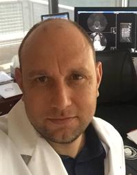
Marcelo D’Abreu, MD
Head of Radiology
Hospital Mae de Deus
© 2026 Medality. All Rights Reserved.
