Training Collections
Library Memberships
On-demand course library with video lectures, expert case reviews, and more
Fellowship Certificate™ Programs
Practice-focused training programs designed to help you gain experience in a specific subspecialty area.
Ultimate Learning Pass
Unlock access to our full Course Library and all self-paced Fellowships.
Continuing Medical Education (State CME)
Complete all of your state CME requirements in one convenient place.
Noon Conference (Free)
Get access to free live lectures, every week, from top radiologists.
Case of the Week (Free)
Get a free weekly case delivered right to your inbox.
Case Crunch: Rapid Case Review (Free)
Register for free live board reviews.
Dr. Resnick's MSK Conference
Learn directly from the MSK Master himself.
Lower Extremities MRI Conference
Musculoskeletal Imaging
For Training Programs
Supplement your training program with case-based learning for residents, registrars, fellows, and more.
For Private Practices
Upskill in high growth, advanced imaging areas.
Compliance
NewTrack, fulfill, and report on all your radiologists' credentialing and licensing requirements.
Emergency Call Prep
Prepare trainees to be on call for the emergency department with this specialized training series.
Training Collections
Library Memberships
On-demand course library with video lectures, expert case reviews, and more
Fellowship Certificate™ Programs
Practice-focused training programs designed to help you gain experience in a specific subspecialty area.
Ultimate Learning Pass
Unlock access to our full Course Library and all self-paced Fellowships.
Continuing Medical Education (State CME)
Complete all of your state CME requirements in one convenient place.
Noon Conference (Free)
Get access to free live lectures, every week, from top radiologists.
Case of the Week (Free)
Get a free weekly case delivered right to your inbox.
Case Crunch: Rapid Case Review (Free)
Register for free live board reviews.
Dr. Resnick's MSK Conference
Learn directly from the MSK Master himself.
Lower Extremities MRI Conference
Musculoskeletal Imaging
For Training Programs
Supplement your training program with case-based learning for residents, registrars, fellows, and more.
For Private Practices
Upskill in high growth, advanced imaging areas.
Compliance
NewTrack, fulfill, and report on all your radiologists' credentialing and licensing requirements.
Emergency Call Prep
Prepare trainees to be on call for the emergency department with this specialized training series.
1 topic, 5 min.
2 topics, 13 min.
35 topics, 2 hr. 11 min.
Morphologic Cirrhosis
5 m.Non-Malignant Nodules in the Cirrhotic Liver
2 m.Dysplastic Nodules
4 m.Portal Hypertension – 2 Patient Case Review
4 m.Pseudocirrhosis
3 m.LI-RADS – Summary
12 m.LI-RADS 1 or 2
3 m.LI-RADS 3
3 m.LI-RADS 3 (THID)
3 m.LI-RADS 4
3 m.LI-RADS 3 Upgraded to LI-RADS 4
4 m.LI-RADS 5
3 m.LI-RADS 5 Change in Lesion Overtime
4 m.LI-RADS 5 – Non-peripheral Washout (NPWO)
3 m.LI-RADS 4 – Psuedo Capsule
4 m.LI-RADS 3 – No Ancillary Imaging Findings
3 m.LI-RADS 5 – Size, Psuedo Capsule, & NPWO
3 m.LI-RADS 5 – 22mm with NPWO
3 m.LI-RADS 4 – Greater than 20mm, Lacking Ancillary Features
4 m.LI-RADS 5 – All-qualifiers, HCC
4 m.LI-RADS – Tumor in Vein (TIV)
6 m.LI-RADS – TIV Infiltrative Tumor
4 m.LI-RADS M – Metastatic Urothelial Cancer
5 m.Fibrolamellar Hepatocellular Carcinoma (FHCC)
6 m.Treatment Options
9 m.Successfully Treated LI-RADS 5
4 m.Successfully treated LI-RADS 5, with Recurrence
4 m.2 Lesions Treated with Transarterial Embolization
4 m.Partially Successful TASE
3 m.Recurrence in a Patient Treated with Radioembolization (Y-90)
4 m.2 Patients with Cholangiocarcinoma
8 m.Hypervascular Mets
4 m.Hypovascular Metastasis from Breast Cancer
4 m.Mucinous Liver Metastasis
4 m.Hypovascular Metastasis Post-op
4 m.0:01
So the following is a patient with
0:02
cirrhosis who presents for screening
0:04
for hepatocellular carcinoma.
0:06
So over here we'll start off by pulling
0:08
up, uh, some of the T1 post-contrast images
0:11
and we'll focus on this lesion over here.
0:13
So here we have a T1-weighted FATSAT
0:16
post-contrast in the arterial phase,
0:19
and this one's similar except we've
0:20
imaged in the portal venous phase.
0:22
And I wanted to use this case to
0:24
showcase what, you know, a LI-RADS
0:26
1 or 2 lesion could look like.
0:29
Many of the LI-RADS 1 or 2 lesions, we've
0:31
already covered in the benign liver mass talk.
0:34
And, you know, they include
0:34
things like cysts and hemangiomas.
0:37
However, this lesion was not
0:38
specifically covered in that talk.
0:40
And it's something we see quite
0:41
often in patients who have cirrhosis.
0:44
So let's look at some of
0:44
the imaging features of it.
0:46
We can see that it's located in the right
0:47
hepatic lobe, probably segment 7 over here.
0:51
And it has, overall, a
0:52
very wedge-shaped look to it.
0:54
Right?
0:55
Straight borders, wedge shape
0:56
at the periphery of the liver.
0:58
It demonstrates arterial hyper-
1:00
enhancement, but on the portal venous
1:02
phase, we really don't see anything.
1:04
It becomes isointense with
1:06
respect to the liver parenchyma.
1:07
Very difficult to see what it is over there.
1:09
And so this combination of this lesion that has
1:12
a relatively geographic distribution, almost
1:15
looks like a little bit of a triangle, very
1:16
sharp linear borders, enhances in the arterial
1:20
phase, you don't see it, is characteristic
1:23
of something called a Transient Hepatic
1:25
Intensity Difference, or a THID for short.
1:30
And this is thought to occur in portions of
1:33
the liver that have relatively poor portal
1:35
flow because of portal hypertension,
1:38
due to liver nodular regeneration.
1:40
So those areas with poor portal
1:42
flow will then compensate and have
1:45
increased arterial hyperenhancement.
1:47
As a result, you see them as these bright
1:50
lesions, relatively geographic in appearance,
1:53
as I said, with sharp and linear borders.
1:58
And, uh, you know, depending on
2:00
your degree of confidence, you
2:02
may call this a LI-RADS 1 lesion.
2:05
You don't need to worry about it.
2:06
You could even call it a
2:06
LI-RADS 2 lesion, potentially.
2:09
Small chance of malignancy, but regardless
2:11
of whether you call it LI-RADS 1 or 2, you
2:13
know, this is the sort of patient you need.
2:14
Who doesn't need specific follow-up
2:16
for this and can return to screening
2:19
back in six months from now.
Interactive Transcript
0:01
So the following is a patient with
0:02
cirrhosis who presents for screening
0:04
for hepatocellular carcinoma.
0:06
So over here we'll start off by pulling
0:08
up, uh, some of the T1 post-contrast images
0:11
and we'll focus on this lesion over here.
0:13
So here we have a T1-weighted FATSAT
0:16
post-contrast in the arterial phase,
0:19
and this one's similar except we've
0:20
imaged in the portal venous phase.
0:22
And I wanted to use this case to
0:24
showcase what, you know, a LI-RADS
0:26
1 or 2 lesion could look like.
0:29
Many of the LI-RADS 1 or 2 lesions, we've
0:31
already covered in the benign liver mass talk.
0:34
And, you know, they include
0:34
things like cysts and hemangiomas.
0:37
However, this lesion was not
0:38
specifically covered in that talk.
0:40
And it's something we see quite
0:41
often in patients who have cirrhosis.
0:44
So let's look at some of
0:44
the imaging features of it.
0:46
We can see that it's located in the right
0:47
hepatic lobe, probably segment 7 over here.
0:51
And it has, overall, a
0:52
very wedge-shaped look to it.
0:54
Right?
0:55
Straight borders, wedge shape
0:56
at the periphery of the liver.
0:58
It demonstrates arterial hyper-
1:00
enhancement, but on the portal venous
1:02
phase, we really don't see anything.
1:04
It becomes isointense with
1:06
respect to the liver parenchyma.
1:07
Very difficult to see what it is over there.
1:09
And so this combination of this lesion that has
1:12
a relatively geographic distribution, almost
1:15
looks like a little bit of a triangle, very
1:16
sharp linear borders, enhances in the arterial
1:20
phase, you don't see it, is characteristic
1:23
of something called a Transient Hepatic
1:25
Intensity Difference, or a THID for short.
1:30
And this is thought to occur in portions of
1:33
the liver that have relatively poor portal
1:35
flow because of portal hypertension,
1:38
due to liver nodular regeneration.
1:40
So those areas with poor portal
1:42
flow will then compensate and have
1:45
increased arterial hyperenhancement.
1:47
As a result, you see them as these bright
1:50
lesions, relatively geographic in appearance,
1:53
as I said, with sharp and linear borders.
1:58
And, uh, you know, depending on
2:00
your degree of confidence, you
2:02
may call this a LI-RADS 1 lesion.
2:05
You don't need to worry about it.
2:06
You could even call it a
2:06
LI-RADS 2 lesion, potentially.
2:09
Small chance of malignancy, but regardless
2:11
of whether you call it LI-RADS 1 or 2, you
2:13
know, this is the sort of patient you need.
2:14
Who doesn't need specific follow-up
2:16
for this and can return to screening
2:19
back in six months from now.
Report
Faculty
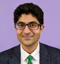
Mahan Mathur, MD
Associate Professor of Radiology & Biomedical Imaging, Vice-Chair of Education & Director of Medical Student Education in Radiology
Yale School of Medicine
Tags
Oncologic Imaging
Non-infectious Inflammatory
Neoplastic
MRI
Liver
Gastrointestinal (GI)
Body
© 2026 Medality. All Rights Reserved.