Neuroimaging of Epilepsy, Dr. Ranliang Hu (10-12-20)
HIDEInteractive Transcript
0:02
Hello and welcome to Noon conferences
0:03
hosted by MRI online.
0:05
In response to changes happening around the world right now
0:08
and the shutting down of in-person events, we've decided
0:10
to provide free daily noon conferences
0:12
to all radiologists worldwide.
0:14
Today we're joined by Dr.
0:16
Ron Hu for a lecture on neuro imaging of epilepsy. Dr.
0:19
Hu is assistant professor of Radiology
0:21
and Imaging Sciences at Emory University,
0:24
associate Program Director of the Neuroradiology Fellowship
0:27
and Director of Stroke Imaging.
0:29
His clinical interest include neurovascular imaging epilepsy
0:32
and functional MRIA reminder, there'll be a q
0:35
and a session at the end of the lecture, so please use the q
0:38
and a feature to ask your questions,
0:39
and we'll get to as many as we can before our Time's Up.
0:42
That being said, thank you all for joining us today.
0:44
I'll let Dr. Hu take it from here.
0:47
Hello everyone again.
0:48
Um, thank you for joining this new conference.
0:51
Uh, my name is Ron Hu.
0:53
I'm from University, uh, Emory University in Atlanta,
0:55
and today I'll be speaking about neuroimaging in epilepsy.
1:01
Um, have I'm involved in a clinical trial on laser ablation
1:05
for temporal lobe epilepsy,
1:06
but have no financial involvement.
1:09
Uh, so the objectives today is to, um,
1:13
talk about why do we image epilepsy, so the rationale
1:15
for imaging, uh, when to image in epilepsy,
1:19
and then go over some of the common, uh,
1:21
pathologies that cause epilepsy.
1:24
Uh, mostly focusing on adult pathologists, uh,
1:27
including temporal lobe abnormalities, malformations
1:29
of cortical development, uh, epileptogenic tumors, uh,
1:33
just a couple of syndromes.
1:34
And then, uh, a, um, kind of a growing kind of a
1:40
pathology, uh, in which is previously underrecognized.
1:44
And then finally, we'll, uh, have some time for questions
1:47
and answers, and please type those into
1:49
the, uh, q and A box.
1:52
So, uh, why do we image in epilepsy?
1:54
Uh, first, let's go over the terminology here.
1:57
So, a seizure is any kind of disturbance,
2:00
electrical activity in the brain.
2:01
And then when you have two unprovoked
2:04
seizures within 24 hours,
2:06
or have one in the, you know, a high likelihood
2:09
of another one that qualifies as a epilepsy syndrome.
2:12
So, epilepsy affects about three
2:15
and a 3.4 million people in the us
2:18
and about the majority of them actually have unknown cause.
2:21
So when patients present initially with seizure, you know,
2:24
we image in order to diagnose an acute cause of seizure,
2:27
such as infection or trauma, intracranial hemorrhage,
2:31
and also to exclude, you know, other, um,
2:33
pathologies like brain tumors.
2:35
Um, so in terms of epilepsy though, uh, we image really
2:40
to kind of guide treatment.
2:41
So, uh, we know that up to a third of patients
2:44
with epilepsy are refractory to medication.
2:47
And nowadays there's, uh, very effective epilepsy surgery
2:51
and even neuromodulatory therapies that can be targeted
2:54
to people with refractory, uh, epilepsy.
2:57
So, and, uh, papers have shown
2:59
that surgical success is actually associated
3:01
with the recession of the focal, uh, seizure zone.
3:05
So being able to identify structural cause
3:07
for focal epilepsy is,
3:09
is very important for treatment planning.
3:13
This is, um, recent abstract,
3:15
I guess it's impressed right now in the H-J-A-C-R, uh, done
3:19
by one of our residents who looked at utilization of imaging
3:22
for seizure and epilepsy in the er.
3:24
We saw that, uh, testing in terms of both EEG, CT
3:28
and MRI for seizure
3:30
and epilepsy has increased considerably between 2006
3:33
and 2014 in the us, uh, going up, uh, you know, 400%
3:38
for MRI in adult patients.
3:41
And this has outpaced the change in number of ED visits.
3:44
And looking at the aggregate data, we found that about nine
3:47
to 12% of, um, these, uh, emergent, uh,
3:51
CT imaging actually found some acute pathology, um,
3:54
in the ed, but in patients with known epilepsy disorder, um,
3:59
43% of them actually had imaging in the er,
4:02
but only 3%
4:03
of those cases actually showed anything that was actionable.
4:06
So, patients with first, you know, uh, new onset CT
4:10
or new onset seizures presented to the ed, you know,
4:13
the ELs about 10% for ct.
4:16
Uh, but then if you have a known epilepsy disorder, uh,
4:19
the yield for imaging in the emergent setting is much less.
4:23
So, um, today I'm gonna focus mostly on CT and MRI.
4:27
Uh, I know, you know, these imaging methods are part of a,
4:31
you know, um, comprehensive workup.
4:33
So that would include pet, maybe even MEEG
4:36
and the invasive monitoring.
4:37
Uh, but for today's lecture,
4:39
we will focus on cross-sectional imaging, CT and MRI.
4:42
Uh, CT is fast
4:43
and available in the first line, uh, imaging tool.
4:47
In the acute setting. It can be used
4:49
for detecting calcifications, uh, such
4:51
as in infectious diseases like neurotic psychosis, uh,
4:55
even cavernous malformations.
4:56
Um, and then osseous defects, uh, for, from trauma
5:00
or, uh, cephaloceles.
5:03
Uh, sometimes CT profusion is done, uh,
5:06
because when they, uh, present,
5:07
we don't know if it's in seizure or stroke.
5:10
So, uh, in the, um, acute setting,
5:13
sometimes CT profusion is done
5:14
and would show hyper profusion in the ictal
5:17
or immediate ictal, uh, period.
5:20
And then finally, uh, we do CT for surgical planning
5:22
and localization, uh, to plan for electro placement, as well
5:26
as, uh, you know, ablations or surgery.
5:29
In terms of, uh, MRI, uh, it definitely is higher costs
5:33
and run into issues with compatibility.
5:35
And a lot of people with, uh,
5:37
epilepsy actually have implanted devices like, uh,
5:40
vagal nerve stimulators and, uh, neuromodulatory devices.
5:44
So compatibility becomes an issue.
5:47
Uh, we typically image patients with epilepsy
5:49
with a dedicated epilepsy protocol,
5:51
preferably at higher strength field strength, like three
5:54
or even nowadays at 70.
5:57
Uh, normally, uh, if the patient is, uh, kind of a older
6:01
or middle age adult, we would give contrast, well,
6:04
brain tumors that are infectious inflammatory processes.
6:08
Um, we sometimes do MR spectroscopy to differentiate
6:10
between the tumor or cortical dysplasia, uh,
6:13
and we sometimes do MR profusion for that as well.
6:16
And then, uh, we, uh, often perform both fm I both task base
6:20
and resting state FMRI for presurgical planning.
6:24
So our protocol here, uh, is, is outlined, uh, to the left.
6:29
Uh, but you know, you can substitute a lot
6:31
of these protocols for, you know, 2D for 3D um, variants.
6:35
So the 2D uh, sequences typically have higher contrast
6:39
and noise ratio, higher in plain resolution,
6:41
but they're thicker slices.
6:43
The 3D um, variants typically have kind of isotropic, uh,
6:48
one millimeter or less resolution
6:49
that you can reformat in any plane,
6:51
but they typically suffer from, uh, a little bit, uh,
6:55
in terms of, uh, signal to noise.
6:58
And when we do a image of hippocampus, it's important
7:01
to do o bleak coronal as opposed to a straight coronal, uh,
7:05
which means that, uh, we orient the, uh, plane
7:07
to be perpendicular to the axis of the hippocampus in order
7:11
to view it in profile.
7:15
In terms of the different causes of epilepsy, in terms
7:18
of structural causes, uh,
7:20
the most common would be mesial temporal sclerosis in adults
7:23
is a focal cause of epilepsy.
7:25
And we also have various tumors.
7:27
Um, this is not an exhaustive list,
7:29
but now any tumor can cause, uh, seizures.
7:33
But these are kind of typically low grade tumors we think of
7:36
as being epilepsy causing tumors, including ganga gliomas,
7:39
DNA and PXA, uh, developmental abnormalities in terms
7:43
of malformations of critical development.
7:45
Uh, various neuro cutaneous, uh, syndromes, uh,
7:49
vascular malformations like cavernous malformations, even
7:52
AVMs sometimes can cause seizures.
7:55
Um, and increasingly recognized, uh, is, uh,
7:58
autoimmune encephalopathy, which I'll show some cases later.
8:01
And of course, any type of infection,
8:03
trauma, and infarction.
8:05
Uh, typically infarction doesn't cause, um,
8:07
seizures in adults, typically.
8:09
That's a presentation in kids.
8:13
So now I'm going to, for the next few minutes,
8:15
focus on temporal lobe epilepsy.
8:17
Uh, first focusing on the imaging appearance
8:20
and, uh, classification of mesial temporal sclerosis.
8:23
Uh, then some of the secondary, uh, kind of, uh, uh,
8:27
imaging features, including amygdala enlargement,
8:30
hippocampal mal rotation,
8:32
and, uh, abnormal temporal lobe signal.
8:34
And then finally, show a couple cases of, uh, autoimmune
8:37
and, uh, infectious encephalitis.
8:41
So, um, kind of going back to neuroanatomy here.
8:44
So what, uh, makes up the medial temporal lobe
8:47
or medial temporal lobe,
8:48
depending on your preference, it's the same thing.
8:51
So basically as three main parts, uh, the amygdala,
8:55
hippocampus, and per hippocampal jars.
8:57
Let's see if this plays here.
9:01
So, um, the amygdala's, you know, anterior portion
9:04
of the medial temporal lobe shaped like an almond.
9:07
Um, and then, uh, you have the onca, which,
9:11
which is the most medial portion of the amygdala.
9:14
Uh, then going back, we start to see the interdis
9:18
of the, uh, hippocampus.
9:19
So this is a hippocampal head,
9:21
which has this characteristic undulating appearance.
9:24
And then as you move back further, uh, you'll, uh,
9:28
see the hippocampal body, uh, which technically kind
9:31
of on the chrono plane begins at the level
9:32
of the red nucleus.
9:34
You can kind of see a faint outline of that there.
9:36
And as you move back further back at the level of the, uh,
9:40
superior inferior ulus, you have the beginning
9:43
of the hippocampal tail.
9:44
And then as you go back further, um, you have the FIA here,
9:48
which you eventually, uh, you know, becomes the fornix
9:52
and kind of loops back forward again.
9:56
Uh, so kind of inferior to the hippocampus.
9:59
We have the, uh, para hippocampal gys, which is here,
10:03
and that's separated from the fusel form gys
10:06
by the collateral sulcus.
10:08
Uh, so this is the collateral sulcus.
10:10
Here we have the gys,
10:13
and then more laterally, we have the superior, middle,
10:16
and inferior, uh, temporal gyri.
10:21
Okay. So, uh, mesial temporal sclerosis, uh, as I mentioned
10:25
before, is the most common cause
10:27
of focal epilepsy in adults.
10:29
Uh, the imaging findings are threefold,
10:31
so you have high signal, which reflects gliosis
10:34
of volume loss, which reflects atrophy
10:37
and loss of internal architecture.
10:39
So the internal architecture, uh, refers to what I mentioned
10:42
before, the, you know, in interdis of the hippocampal head,
10:47
but also this kind of, uh, strip you visualize on most MRIs,
10:50
especially three T or above.
10:52
You'll see this kind of thin linear, uh,
10:55
dark signal on T two, which we is a white matter tract.
10:58
So that's the stratum radi atom, that's a part
11:01
of the white matter tract that goes into the hippocampus.
11:03
So you should be able to recognize that, uh,
11:06
on any good quality MRI, uh,
11:08
better in three T than in 1.5 T.
11:11
However, even with, you know, current imaging techniques,
11:14
you know, 20 to 30% of, uh,
11:16
MTS can actually be m MRI negative.
11:19
So, um, there are papers that show that, you know,
11:22
in these MRI negative cases 70 can be helpful.
11:26
And, and we know that from a surgical literature,
11:29
that the postoperative outcome correlates with the kind
11:32
of degree of abnormality and MRI.
11:34
So if you have a definite MTS, you know,
11:36
the surgical outcome for treating
11:38
that is, is gonna be better.
11:41
There is a classification system for MTS, which, you know,
11:44
we don't really get into, uh, as radiologists, it's more
11:47
of a pathologic, uh, subtype, uh, system.
11:50
The type one is the most common,
11:51
which is neuronal cell loss,
11:53
and gliosis predominantly involving the ca one
11:56
and ca four regions.
11:57
Um, so here and here and type two and three are less common.
12:02
Uh, type two is a ca one predominant neuro node loss,
12:05
so more kind of the lateral portion of the hippocampus.
12:08
And then C uh, type three is a ca four,
12:11
and D jar is predominant volume loss.
12:16
Uh, so this is an example of what I showed before.
12:19
So, uh, typically, um, I like to use the T two
12:22
to look at the kind of volume, um,
12:25
and the internal ex architecture.
12:27
So here on the, uh, left side here, lemme just change
12:31
to the laser pointer.
12:33
So Le left side of the patient, we have, uh, atrophy
12:38
of this hippocampus with loss of an internal architecture.
12:41
And then the flare shows that it's hyperintensity signal.
12:47
So there are various secondary findings of, uh,
12:51
mesial temporal sclerosis, uh,
12:53
enlargement of the temporal horn.
12:55
Uh, you can have atrophy of the perim capal jars, widening
12:59
of the collateral sulcus atrophy of the,
13:02
its later mammary body here, uh, as well as, you know,
13:05
the whole, uh, pape circuit.
13:06
So the fornix, uh, even involving the thalamus.
13:10
Uh, you can also have either increasing
13:13
or atrophy of the amygdala, which is a, uh, kind
13:15
of an under-recognized sign, no show later.
13:20
Uh, often, uh, we have dual pathology, which means
13:24
that there is, uh, mesiotemporal sclerosis,
13:26
but it's actually secondary to a different, uh, kind
13:30
of a seizure, uh, provoking, uh, pathology.
13:34
So it occurs about 15% of cases.
13:37
This is a case of the left MTS that actually had a, uh, uh,
13:42
focal cortical dysplasia.
13:47
This is a, uh, case of, um, MTS on the left,
13:50
but there's an underlying, uh, kind
13:52
of a developmental venous
13:54
or, uh, underlying, uh, cavernous malformation.
13:57
Here you see the complete hemo citrin rim of the cab mount,
14:00
and you see associated DVA, um, beating, uh,
14:04
away from that cab mount.
14:05
So it's important when you look at MTS
14:07
and not get kind of, uh, satisfaction of surge
14:10
and stuff right there, it's important to kind of look
14:12
through the entire temporal lobe, even outside
14:14
of the temporal lobe, to look for dual pathology,
14:17
because it really affects treatment.
14:21
Um, sometimes you can get bilateral MTS,
14:24
so it's observed in about three to 10% of cases.
14:27
Uh, so here's an example
14:28
where you have atrophy in hyperintense signal
14:32
of both hippocampi.
14:34
Uh, some usually though the symptoms,
14:36
you know, uh, is unilateral.
14:38
So, um, you know, one of these is, uh, is, uh,
14:43
causing the seizures, but it's very hard to tell by imaging.
14:45
So, uh, they'll probably need more invasive, uh, testing.
14:51
Nowadays, I think a lot of institutions have started using
14:54
kind of volumetric analysis, uh,
14:57
to assist in quantifying the, uh, hippocampal size.
15:00
And here we use, uh, a neuro con,
15:02
but there are others available.
15:04
And there was a paper actually looking at, you know,
15:06
comparing the sensitivity
15:08
and specificity of, uh, neuroco versus neuroradiologist.
15:11
And it was, it was actually neuro con one,
15:13
but it was statistically not significant.
15:15
Um, but this is only looking at volume loss.
15:18
So, uh, it's important for us as radiologists to kind
15:21
of check the neuro con for any type
15:23
of quantitative analysis, make sure the segmentation is
15:25
correct, and to also, um, correlate
15:28
with other imaging findings in clinical history.
15:32
And, uh, the normal, uh, usually these kind
15:34
of software spits out a number, which is asymmetric index.
15:37
It's basically, um, uh, the percentage kind
15:42
of asymmetry between the two sides.
15:44
And, uh, in normal, uh, subjects, you can expect an index
15:49
of 2.1%.
15:51
So there is some natural variation in terms of size
15:53
of the hippocampus, and it's important not
15:55
to call these too tightly.
16:00
I mentioned, uh, 70 T before.
16:03
Um, there, this is a paper I actually know pretty old now,
16:06
nine years ago, that, um, looked at 70, uh, in MTS.
16:11
And, uh, you know, 17 is becoming more popular.
16:14
There's clinically approved, uh,
16:16
devices now from the major vendors.
16:18
So we'll beginning to see this more often.
16:21
Uh, in terms of the, uh, what you can see,
16:23
you can see the interdis very clearly.
16:25
You see the striation much more clearly on seven
16:28
T than on three T.
16:30
Um, in this group of, um, a small group,
16:32
they actually observed hippocampal mal rotation in
16:35
four out of 11 patients.
16:37
And I'll, I'll mention what that is later.
16:40
Uh, in, in temporal lobe epilepsy, they found that there was
16:43
posity of the interdis of the, uh, hippocampal head in all
16:47
of their patients with, uh, temporal lobe epilepsy.
16:50
Uh, even they saw that in three out
16:52
of eight on the contralateral side,
16:55
and they saw, they saw a loss of that striation
17:00
in terms of hippocampal marrow rotation, basically
17:02
what it is, is a, uh, it's, uh, kind
17:05
of a abnormal morphology of the, uh, hippocampus
17:09
where normally it's kind of lying on its side,
17:12
but in the abnormal, uh, side, it's kind of more upright.
17:16
And as a kind of a, a secondary feature,
17:19
the collateral sulcus, which is normally more kind
17:22
of at a 45 degree angle, it's more, uh, upright as well.
17:27
You have abnormal vertical and deep collateral sulcus.
17:30
Um, there's some debate, uh, in, in terms of the cause
17:34
of relationship between, uh, this finding and seizure.
17:37
Uh, I think most people now believe the
17:39
CBA developmental variant.
17:41
Uh, it's not directly related to mts,
17:45
but can be a sign of kind of a general developmental, um,
17:50
abnormality, you know, uh,
17:51
that signals something went wrong in the, uh, formation
17:54
of the temporal lobe, but it's not directly related to MTS.
18:01
Uh, Amy, the amygdala enlargement is something
18:04
that's also kind of recently recognized, um,
18:07
as a part of MTS.
18:08
So, um, you can have a am amygdala enlargement in
18:12
oscillation, uh, with increased signal, um,
18:15
and on pathology, you can have, basically, it shows, uh,
18:19
a type of dysplasia with hypertrophic neurons
18:22
and atrophy, uh, gliosis, uh, often though, uh,
18:25
in intraoperative recordings, even though the, uh,
18:29
findings in amygdala, they also record
18:31
that the seizure is actually coming from the hippocampus.
18:34
So, um, it's kind of a
18:37
unknown whether this is a secondary finding of, uh, MTS
18:41
or a primary cause of, uh, you know, distinct type
18:44
of mesial Temporal sclerosis,
18:48
reportedly it's less associated with febrile seizures
18:50
and has a later onset than the classic MTS
18:53
that's only involved in the hippocampus.
18:56
Uh, important to recognize though there, uh, kind of mimics
18:59
of, uh, this, uh, amygdala enlargement, uh, in the immediate
19:03
kind of postictal period, you can have enlargement of the,
19:06
uh, amygdala in the hippocampus.
19:09
Uh, you can also have low gliomas that occur in this region,
19:13
uh, and, uh, even encephalitis.
19:15
So, um, kind of important to recognize those as well.
19:21
Anterior temporal lobe signal is, uh, is another kind
19:24
of subtle secondary feature that you may observe, um,
19:28
is reportedly observed, uh,
19:30
in the ipsilateral anterior temporal white matter in the
19:33
third of patients with, uh, me,
19:35
medically refractory temporal lobe epilepsy.
19:37
And what it is basically is kind of this, uh,
19:40
hypertense signal in the white matter in the, uh,
19:43
temporal pole, or adjacent to the hippocampus in the,
19:46
in the collateral white matter.
19:48
And you can visualize better, uh,
19:50
if your institution does a, uh, kind of a,
19:52
a double inversion recovery.
19:53
So we do a double inversion recovery sequence here,
19:56
which kind of has two saturation pulses, saturates the, uh,
20:00
CSF, as well as, uh, the, uh, my kind
20:03
of the myelin in order to accentuate the, uh,
20:06
abnormalities in a white matter.
20:08
So DIR tend to be more accurate, uh,
20:10
to lateralize this finding.
20:14
This is, uh, an example of, uh, autoimmune encephalitis.
20:17
Basically, you have kind
20:19
of the enlargement in abnormal signal of the, uh,
20:22
both the left and right, uh, ma, the left and hippocampus.
20:26
Um, these, uh, you know,
20:28
tend can have some left menal enhancement if the image these
20:31
over time, they tend to develop atrophy
20:34
and scoliosis over time.
20:36
And as you know, you know, there are various causes
20:37
of autoimmune encephalitis, uh,
20:40
vari various antibodies like the anti NMDA receptor
20:44
antibody, as well as multiple tumor associated
20:47
autoimmune causes.
20:51
This is a case, um, of a infectious, um, uh,
20:56
temporal lobe seizure.
20:58
Uh, it's actually presented to us,
21:00
you know, as a stroke alert.
21:02
So the patient, uh, went through this whole C-T-C-T-A
21:06
and CTP, and on the CTA we saw there was some hypo density
21:10
in the right insula, uh, even sub insular white matter.
21:15
And the, uh, preliminary report was
21:17
that this may be a right MCA stroke,
21:19
but if you kind of, uh, look more closely,
21:22
there's actually hypo density also involved the right
21:25
amygdala, which is in a different vascular territory.
21:28
So the amygdala in the very tip
21:30
of the hippocampal head is actually supplied
21:33
by the anterior choroidal artery,
21:34
which arises from the ICA is not from the MCA.
21:38
And then in vascular imaging,
21:39
we saw there was no large vessel occlusion.
21:41
So the ICA was not occluded.
21:43
The MCA was widely open,
21:45
and there's actually kind of a increased, uh, prominence
21:49
of the kind of particular branches on the right.
21:51
And on profusion there was increased blood volume
21:53
and increased, uh, blood flow.
21:56
So this was a case of herpes encephalitis, uh,
21:59
which has shown better on MRI.
22:01
You see, the extent
22:02
of the abnormal signals is more kind of broad.
22:05
It involves the frontal lobe, temporal pole, and insula.
22:10
There was associated left meningeal enhancement.
22:12
There was cortical restrict diffusion
22:14
that reflects probably recent seizure activity.
22:17
And if you pay attention even more closely,
22:20
there is layering, uh, you know,
22:23
material within the occipital horn
22:25
that could be biogenic material or some blood product.
22:28
So, um, you know, uh, we should always be aware
22:31
of these kind of, uh, pathologies,
22:34
even when they present us, is not seizures
22:36
'cause uh, the treatment is, uh, is very different.
22:41
So, moving on to, uh, malformations of cortical development,
22:45
um, this is a pretty broad, uh, category of, uh,
22:49
diseases which can, um, you know, uh, result from the rest
22:53
of any, uh, step of the normal, uh, kind of, uh, migration
22:58
and, uh, organization of, uh, uh, neurons
23:02
as they migrate away from the germinal
23:04
matrix toward the cortex.
23:06
So when you have abnormal proliferation
23:08
or apoptosis, you can develop microcephaly, heme, cephalic,
23:12
or type two cortical dysplasia.
23:14
Uh, if you have a rest of dys normal, uh, migration,
23:18
you can get heterotopic, uh, gray matter.
23:21
Uh, so basically you can get per ventricular, uh,
23:25
or subdermal heterotopia.
23:27
You can get, uh, subcortical heterotopia or lissencephaly
23:31
or smooth brain.
23:33
And then finally, when you have abnormal cortical
23:35
organization, uh, you have, you can develop poly micro jia,
23:39
uh, sli, and for some reason, type one
23:42
and three FCD, uh, are funded under a category
23:46
of abnormal cortical organization.
23:48
Uh, but type two is abnormal apoptosis.
23:54
So, uh, focal cord dysplasia,
23:56
there is actually three types as well.
23:58
Uh, the type that we usually visualize is type two, uh,
24:02
specifically type two B, so dysmorphic
24:04
neurons with balloon cells.
24:06
But, um, you know,
24:07
type one you can see some imaging findings.
24:09
So type one refers to, uh,
24:12
just abnormal cortical lamination.
24:14
Uh, on imaging, you'll see some subtle blurring
24:17
of gray white junction, uh, but less so than type two.
24:20
You can have kind of segmental
24:22
or lower atrophy with loss
24:24
of the regional white matter volume.
24:26
Uh, you can moderate increase in T two signal
24:28
in the underlying white matter.
24:30
Uh, so these can be subtle and can be missed on imaging.
24:33
Uh, type two, uh, is the one,
24:35
typically type two B is the most common one that we see, uh,
24:38
is more common to be, uh,
24:40
more commonly found in frontal than temporal lobe.
24:45
And what you see is a cortical thickening, uh,
24:47
with increased T two signal.
24:49
There's marked blurring of the gray white junction,
24:51
and sometimes you see this, uh, trans mantal sign
24:54
of abnormal kind of white matter, um, underlying the,
24:57
uh, cortical dysplasia.
24:58
And type three is basically a, uh, SCD that's associated
25:02
with other pathologies.
25:07
So, um, this is an example of a, uh,
25:09
type two focal cortical dysplasia.
25:11
Um, this is a kind
25:13
of an abnormal gray white differentiation,
25:15
cortical thickening in the right temporal lobe.
25:17
That's important to kind
25:18
of look at these in different planes
25:20
because you don't wanna be confused by volume averaging, um,
25:24
when you look at any single plane.
25:25
So here, this one proves to be real on both planes.
25:29
And then on a T two, you can really appreciate the kind
25:32
of the cortical thickening
25:33
and even kind of abnormal signal in
25:35
the adjacent white matter.
25:38
And pathologically, this is very similar to
25:40
what you see on the histology.
25:42
Uh, you see cortical thickening
25:44
and blurring at the gray white junction.
25:48
This is, uh, an example of a transman sign.
25:51
So focal cortical dysplasia here, paramedian, uh,
25:54
it looks like frontal lobe.
25:56
Um, you have kind of increased signal in, in a blurring
26:00
of the gray white differentiation here.
26:02
Uh, but then you also see this kind of band
26:04
of abnormal white matter extending
26:06
toward the ventricle margin.
26:08
And this is, uh, the trans mantle sign.
26:10
And you can, uh, see this also better on the double inver
26:13
copy sequence at 70 T.
26:17
Uh, you can see a lot better than at three T as expected.
26:22
Uh, this was a paper, uh, that looked at 21 patients with,
26:25
uh, intractable epilepsy with negative 1.5
26:29
or three T studies,
26:30
and they found that six of these 21 patients were found
26:34
to have FCT on MRI confirmed by pathology at 70.
26:38
Um, so on the Mr I 70,
26:41
you basically can see much
26:42
better at the gray white differentiation.
26:43
Here you see this kind of abnormal, uh,
26:46
lamination gray white loss here,
26:49
which you cannot appreciate at three T.
26:54
Um, just to complete the picture.
26:56
So this is what Hemi, omega
26:57
and Celi looks like, not to be confused with hemi atrophy.
27:00
So when you look at these cases, always is important to kind
27:04
of think about which side is abnormal here.
27:06
So in this case, the right side is abnormal.
27:09
Uh, we have actually enlargement of ifs lateral, uh,
27:13
uh, ventricle here.
27:14
So, um, this is a type of, uh, this, a example
27:18
of a Hemi Omega cephalic encephalopathy.
27:22
So al heterotopia, basically, you can have a rest
27:25
of gray matter, uh, migration anywhere
27:28
between the ventricle and the cortex.
27:30
So when this occurs in the, uh,
27:32
ventricle sub al heterotopia here, we have extensive kind
27:35
of angulating abnormal, uh, gray matter.
27:37
So these should follow, uh,
27:39
gray matter signal all sequences.
27:41
So on the T two, it should look just like the cortex.
27:44
And then pure rage, same intensity as the cortex
27:48
and flare, uh, usually should not have hyperintense signal.
27:53
This is a case of a band heterotopia.
27:55
So these neuro made it out a little further
27:58
that got arrested, kind of in the deep white manner.
28:05
And this is an example of a closed lip schizo celi.
28:08
Uh, these are always lined with abnormal poly MicroAge area.
28:12
So the cleft is, uh, lined by poly MicroAge area.
28:19
Let's see, 30 minutes.
28:22
Um, so, uh, now we're gonna kind
28:26
of focus more shifting gears toward the tumors.
28:29
Uh, going from benign to, uh, more high grade.
28:32
Of course, you know, higher grade, like grade three
28:35
and grade four tumors can cause seizures.
28:37
Uh, but we don't typically, you know, they're not epilepsy,
28:40
uh, causing tumors
28:41
because, uh, you know, they're their own entity
28:44
and are treated more aggressively.
28:46
So in terms of the, uh, more lower grade tumors,
28:49
we can talk about hematomas, which are basically arrests
28:53
or basically normal tissue and an abnormal place.
28:56
Uh, there's this new entity called multinodular evacuating
28:59
neuronal tumor, which I'll show, um,
29:02
which can cause seizures.
29:04
Uh, the classic kind of, uh, lower grade tumors
29:07
that cause seizures would be the dnet glioma, uh, PXA
29:12
and, uh, oligo dro gliomas are also common type
29:16
of tumors that can cause seizures.
29:19
So this multinodular evacuating neuronal tumor is, um,
29:23
is a recently recognized entity.
29:25
Um, pathology still don't agree,
29:27
whether it's a cy architectural pattern versus
29:30
a benign tumor.
29:32
What it looks like basically is like if you splash paint,
29:35
you know, in a subcortical white matter,
29:37
and it just, it always looks like this.
29:39
So almost like looks like prominent perivascular spaces, uh,
29:43
but they're more focal.
29:45
So they occur in the subcortical white matter.
29:47
They typically have no enhancement or mass effect.
29:50
Uh, they can be an incidental finding without any symptoms,
29:54
or they can be associated with seizures.
29:58
Hematomas, as I said before, are basically normal tissue
30:02
and abnormal place, so they're overgrowth of tissue.
30:06
Um, the classic one for boards
30:08
that we all study is the tumor cerium hematoma.
30:11
Uh, the tumor cerium is kind of like the floor
30:14
of the third ventricle, part of the hypothalamus,
30:16
but you can get hypothalamic gliomas
30:19
or, um, kind of anywhere.
30:22
Uh, they can be sessile or pedunculated.
30:24
The tumor scenario one is associated with seizures.
30:28
Uh, they should follow kind of gray matter on all sequences.
30:31
Sometimes they can have a little bit of gliosis,
30:33
so they may look a little bright on flare,
30:35
but mostly they should follow gray matter on the, uh,
30:39
all sequences, and they don't have any
30:40
enhancement or calcification.
30:44
Uh, it's important to kind of, uh,
30:47
differentiate these from hypothalamic gliomas,
30:50
which are actual tumors,
30:52
even though they might be low grade, uh,
30:54
they have neoplastic potential.
30:55
So the gliomas, they tend to be more T two hyperintense.
30:59
Uh, they tend to be more expansive.
31:01
Here we have one, uh, kind
31:03
of here on the T two weighted image.
31:05
You can almost, you know, miss it
31:07
because it's so, T two hyperintense almost blending
31:09
into the rest of the ventricle.
31:11
It's actually on the kind of fiesta, uh, case sequences.
31:14
You can really visualize, uh, this tumor in its mass effect.
31:19
These may or may not have enhancement.
31:21
Uh, typically in kids, there's going to, these are gonna be,
31:24
uh, pilocytic, astrocytomas and associated with NF one.
31:30
There's another example of, uh, emmic,
31:33
oh, this is actually the same case.
31:34
This is a pre T one.
31:36
Uh, here, post T one contrast, you see a little bit
31:39
of enhancement, uh, in here.
31:41
So having enhancement
31:42
or not, as you know, doesn't mean it's a high grade.
31:44
So pilocytic astrocytoma typically have enhancement in
31:48
their, uh, low grade tumor.
31:53
Uh, so now begin kind of to talk about the classic kind of
31:58
with the nodule type of tumors.
31:59
Uh, well, not yet.
32:01
This dnet is actually, uh, you know, typically, uh,
32:05
for board purposes, a bubbly corly based lesion.
32:08
Uh, it's, uh, type one,
32:09
it's a grade one glio neuronal tumor has this kind
32:12
of classic bubbly appearance.
32:14
So almost like a bunch of, uh, soap bubbles.
32:16
Uh, they are more commonly found in the temporal lobe, less
32:20
so in the frontal lobe.
32:21
They rarely enhance
32:22
or calcify, which is important to recognize.
32:25
Uh, when you kind of compare it to the other, uh,
32:28
things I'll show later, uh,
32:30
because they're very slow growing, if they're kind
32:33
of near the cortex,
32:34
they can actually scout the adjacent calvarium over time.
32:38
So important to kind of look at a ct if you have CT
32:41
or even on the MRI to see if you have that finding.
32:44
And that that's a feature that you'll see in a lot
32:46
of slow growing, uh, lesions.
32:48
But you can see that in dnet gang gliomas are,
32:53
uh, also type one glio neuronal tumors.
32:56
Uh, these are the classic kinda assist
32:59
with the nodule, uh, tumors.
33:01
Um, they have variable enhancement they tend to enhance, uh,
33:06
a lot more often than DAD nets don't, uh, commonly enhance.
33:10
And you tend to have, uh, more classification
33:12
with gang glioma.
33:16
Um, pleomorphic ex santo astrocytoma
33:19
or PXA are slightly higher grade.
33:21
They're grade two astrocytomas.
33:23
Uh, they also have this cyst, uh,
33:26
with enhancing nodule uh, appearance.
33:29
They tend to have a little more edema than the gang gliomas.
33:32
And, uh, they can even have a little bit
33:34
of a lepto menal enhancement.
33:36
Sometimes you'll see a little bit of, uh,
33:39
enhancement in the, uh, overlying lepto meninges.
33:45
This is not a, uh, interactive session,
33:47
so I can't have you guys answer questions.
33:49
So, th this is a caven smell formation.
33:51
So you can see this kind of, uh, susceptibility artifact,
33:54
T two, uh, dark, uh, lesion here
33:57
with some underlying white matter abnormality in some kind
34:01
of, uh, peripheral kind of sinusoidal kind of enhancement.
34:05
So this is a cavernous malformation.
34:11
So this is, uh, uh, a lesion in the left frontal lobe.
34:15
I guess you can feel free to put in the text what you think.
34:19
This is tumor or cortical dysplasia.
34:22
Um, but this turned out to be a tumor.
34:25
Uh, so this is a low grade tumor because it's expansile.
34:30
Um, we, uh, there was no significant enhancement,
34:33
um, and we did Mr.
34:35
Or Mr. Profusion and showed
34:36
that this was a hypo, uh, perfusion.
34:39
So, low grade tumors, uh,
34:41
typically don't have elevated perfusion.
34:44
So, uh, it doesn't, having kind of normal
34:47
or low perfusion doesn't exclude low grade tumor.
34:50
So this tend to, this, uh, turned out
34:52
to be oligo dro glioma.
34:55
Sometimes we do spectroscopy.
34:58
Uh, they try to differentiate between low-grade tumor
35:01
and cortical dysplasia.
35:03
And, uh, a normal MRS spectrum looks kind of like this.
35:06
Um, you know, this one didn't have the traditional hunter
35:09
peak, where you have the choline creatine,
35:10
and NNA, um, we see this sometimes, um,
35:14
doesn't mean it's abnormal.
35:16
Here we have an example of a focal cortical dysplasia.
35:21
Now, the problem with, uh, MRS is that everything causes,
35:26
um, increased choline decrease in a, so in this case,
35:29
the focal dysplasia, cortical dysplasia increased choline
35:33
because of, uh, increased membrane turnover, uh,
35:35
decreased NAA,
35:37
but it does that in a much lesser degree than the tumor.
35:41
So in the tumor, you have a greater increase in choline
35:45
and a much greater decrease in n na.
35:48
So, uh, papers have shown, if you look at the, uh,
35:50
the degree of, uh, the,
35:54
not just the direction of change,
35:56
but the actual magnitude of the change, you can kind
35:59
of differentiate between low grade tumors
36:01
and focal cortical dysplasias.
36:03
In practice, though, I think, uh, most people
36:06
who look at MMRS, uh, tend to agree
36:09
that these are not clear cut.
36:11
So there's no, um, universally accepted thresholds
36:15
for separating these things.
36:17
So it's kind of, you have to look at the lesion as a whole
36:20
and use your clinical
36:21
judgment checking on time.
36:27
Um, so we have, uh, 20 minutes left.
36:29
I'll go over just a couple of kind of syndromes
36:32
that you may see in adult neuro imaging.
36:35
Um, there's a bunch of pediatric epilepsy syndromes, which,
36:39
you know, I won't talk about today.
36:41
And then I'll focus a little bit on cephaloceles,
36:43
which are increasingly recognized as causes
36:46
of seizure or epilepsy.
36:49
So this is a case of a Sturge Weber syndrome.
36:52
Uh, you can see kind of calcifications.
36:55
Uh, so these, these are tend to to be childhood.
36:57
They're recognized in childhood, so usually
37:00
by the time they're adults, they, they should know
37:01
that they have s webber.
37:03
But you may see this, um, uh, basically calcifications.
37:06
You have, uh, lepto meningeal enhancement, um, due to the,
37:10
uh, abnormal kind of basically venous, uh,
37:13
drainage in a po angio mitosis that occurs in, uh, s Weber.
37:17
And they usually will have some kind of port wine stain
37:20
or, uh, facial, um, kind of, uh, uh,
37:24
stigmata on the ipsilateral side.
37:29
This is an example of a d**e Davidson Meison syndrome.
37:33
Basically, it's a cerebral hemi atrophy,
37:36
and this is more description of a constellation
37:39
of findings in the clinical syndrome rather than an entity.
37:42
So, uh, various things can cause this, uh, can be from kind
37:46
of a early insult, uh, some kind of encephalitis or,
37:50
or idiopathic.
37:51
So what you see is basically, uh, atrophy, uh, of one side
37:56
of the brain, as we can see here.
37:58
Um, surprisingly,
38:00
there's not much gliosis given the degree of atrophy.
38:02
So you know that the insult probably occurred very early.
38:05
Uh, you may see kind of thickening
38:07
of the underlying calvarium.
38:08
So here, uh, in kind of indicating that this is a kind
38:12
of a slow growing, slow, uh, process
38:15
that the calver actually thickened to fill that empty space.
38:18
And you can even see kind of, in this case,
38:20
we don't have it, but you can see kind of enlargement
38:23
of ipsilateral, uh, perinasal sinuses.
38:26
And here we have kind of more ln degeneration
38:29
of the ipsilateral ce.
38:31
Uh, this is a little oblique,
38:33
but you can also see kind of just the, the middle
38:36
of cranial fossa to be, uh, smaller on that side.
38:39
So th this is a clinical syndrome
38:41
that can be from various etiologies
38:43
or, um, the idiopathic, um,
38:50
switching gears to encephalocele.
38:52
So, uh, ence seals are basically protrusions
38:57
of brain through, uh, a defect in the, uh, in the calvarium.
39:02
So, uh, it can be a natural defect such as the FMO Valley.
39:06
So in patients with idiopathic intracranial hypertension,
39:10
because of the chronic elevated intracranial pressure,
39:13
you can actually have kind of almost a erosion
39:15
of the FMO valley here demonstrated on the
39:17
3D reconstruction.
39:19
So it gets larger and part
39:21
of the basal temporal lobe actually herniates through.
39:24
And sometimes on flare
39:25
or, you know, DIR,
39:26
you can actually see gliosis in that portion of the brain.
39:30
So now, in these patients, uh,
39:32
de encerios can be a seizure focus.
39:35
So they can occur, uh, in multiple different locations,
39:39
in this case, through a natural foramen.
39:42
But other times you can have these kind
39:44
of thoro osseous defects also as sequela of ih, uh,
39:48
basically have a, almost like a chronic pressure erosion
39:51
of the inner table of the area.
39:54
And you have this herniation
39:56
of the temporal pole into that area.
39:58
And you can even see there's xva dilatation of that, uh,
40:02
CSF space there, indicating that there's almost kind
40:05
of a tenting or, uh, traction of this temporal pole.
40:09
It's not just like, you know, freely going in there,
40:12
it's actually being pulled or stuck in there.
40:14
And this, uh, it was, uh, studied with a stereotactic EEG
40:18
and found to be the seizure source.
40:20
So, uh, these, uh, occur very commonly where I practice,
40:25
where there, you know, there's a lot of obesity, uh, a lot
40:28
of patients with ih.
40:30
Uh, and you know, up until a few years ago,
40:33
these were under recognized.
40:34
And, uh, you know, the patients were just, uh, you know,
40:39
thought to have just general, just un nonfocal epilepsy
40:43
or, uh, without a structural cause.
40:45
But they actually have this cause
40:47
and then they can be, uh, studied.
40:49
And these, um, areas can either be repaired
40:52
or actually, um, one
40:54
of our neurosurgeons does a laser ablation
40:56
of these, uh, areas.
40:58
So, uh, sometimes though, uh, patients with IH
41:02
commonly will have bilateral encephalocele,
41:04
so it can actually be sometimes difficult to determine which
41:08
encephalocele, uh, these are from.
41:11
So, uh, they won't be studied usually
41:13
with a stereotactic EEG to confirm.
41:17
Uh, ence seals can also occur elsewhere.
41:20
Uh, I've seen them, you know, in weird places,
41:22
even transverse sinus.
41:23
You can have herniation
41:24
of the brain into the transverse sinus.
41:26
In this case, uh, it's a frontal sinus, uh, ence seal.
41:31
Uh, this patient was, uh, actually admitted as an inpatient
41:35
with, uh, a meningitis and had this brain MRI,
41:39
and it looks initially on first glance
41:41
to be, you know, sinusitis.
41:43
But if you look closer, there's actually a discontinuity
41:46
of inner table of the calvarium.
41:48
On the coronal.
41:50
You actually see this gyro form
41:51
or cerebral form appearance of that tissue.
41:54
It's not just, uh, sinus disease.
41:56
And on the CTA, we actually saw vessels going into it.
42:00
So, which really cinched the deal for us
42:02
that this was an encephalocele.
42:04
And this patient actually has epilepsy, um,
42:07
from chronic meningitis.
42:09
They, they, they admitted multiple times in the past
42:12
with meningitis and had imaging,
42:14
and it was always, you know, called sinusitis.
42:17
And that was under recognized to be encephalocele.
42:23
So, uh, in conclusion, imaging is important
42:25
to detect treatable causes of epilepsy.
42:27
That's very important for, uh, treatment, uh, triage,
42:32
as well as kind of predicting the outcome.
42:35
Uh, the imaging finding can be very subtle.
42:38
So it's important to use kind
42:39
of high field MRI optimized techniques, uh, especially
42:43
to detect, uh, mesial temporal sclerosis
42:45
and focal cortical dysplasias.
42:47
And also, it's important to, you know, at least, you know,
42:50
attend seizure conference
42:51
and see how, um, the, the team, the epilepsy team kind
42:56
of uses all the information, uh, with PET and semiology
43:00
and, uh, the monitoring, uh, to come up the diagnosis.
43:04
Encephalocele are, uh, pretty common cause of FFC,
43:08
at least in some parts of the world where IH is more common.
43:11
And, uh, it's important to recognize those, uh, as well.
43:17
So with that, I'd like to thank you for your attention.
43:19
Uh, here's my email address on the left,
43:22
and you can also contact me via Twitter.
43:24
Um, we are having an open house
43:27
for our neuroradiology fellowship at, at end of the month.
43:29
So, uh, everyone who's, uh, a resident is welcome to join
43:33
that and learn more about, uh, neuroradiology at Emory.
43:37
And with that, uh, I'll start to answer some questions.
43:42
Okay, so someone asked no routine a SL to look
43:46
for Interictal Hyperperfusion.
43:48
Ictal hyperperfusion.
43:51
Okay, so, um, we don't typically do a SL, uh,
43:55
for epilepsy protocols.
43:57
Um, we usually don't image, uh,
44:01
with MRI in the ictal stage.
44:03
So, um, typically we're imaging in the interictal stage.
44:08
There are papers that say that in the interictal stage, uh,
44:12
you can use a SL almost like a pet,
44:14
you'll see, uh, hypoperfusion.
44:16
But, um, that is not part of our protocol.
44:19
Uh, most of our patients do get, uh, pet imaging as well.
44:22
So, uh, that that is true, that, uh,
44:25
you can use a SL in the same way,
44:27
but I don't think it's commonly used.
44:33
When do you refer patients for PET ct?
44:37
Um, so, uh, in our con in,
44:41
I guess the practice will be different, uh,
44:43
depending on where you practice.
44:45
At our center, um, most patients will get a pet.
44:48
Uh, other places, you know, you may have, um, patients who,
44:52
you know, who have discordant imaging findings
44:55
with their clinical feature or some kind of
44:57
more atypical cases, they'll get pet.
44:59
But at our center, um, it's part
45:01
of our standard epilepsy protocol for refractive epilepsy.
45:05
They'll get PET imaging. Um, as part of that workup.
45:13
Another question, isn't enhancing gliomas
45:17
or aren't enhancing gliomas as malignant?
45:20
Um, not necessarily.
45:21
So in the adult population, uh,
45:26
I guess you can phrase it differently.
45:28
Um, so non enhancing tumors
45:33
are typically lower grade,
45:35
but having enhancement doesn't
45:37
really mean it's higher grade.
45:39
Um, so typically when you see a non enhancing glioma,
45:42
typically you're talking about low grade
45:44
glioma, but that's not always true.
45:45
You can't have, you know, um, you know, types
45:48
or WHO uh, grade three tumors that don't enhance
45:53
or have minimal enhancement.
45:56
But, uh, some tumors like pilocytic astrocytomas
46:00
or px, a, even like gliomas, even D nets,
46:03
they can have enhancement and they're not malignant.
46:11
So click here, click here
46:14
in Ictal PET or Interictal pet.
46:17
So typically, uh, PET is done interictal,
46:22
uh, because just the, you know, the logistics of, uh,
46:25
doing the PET with, uh, preparing the FDG
46:28
and everything, um, we do sometimes do spec, uh,
46:33
with ictal and interictal spect
46:35
and obtain the subtraction image.
46:38
Um, so we typically do ictal spect, but not ictal pet.
46:45
Someone asked, when is MEG useful?
46:48
Uh, MEG uh, is also an adjunct to,
46:52
so it can be part of a comprehensive epilepsy workup.
46:56
Uh, MEG is not always available.
46:58
It's actually, you know, only available at a few centers.
47:01
So when there is, uh, kind of atypical findings, uh,
47:05
sometimes, uh, we will refer patients for MEEG
47:09
and then, you know, uh, so it is a discussion
47:12
that usually occurs in the epilepsy conference.
47:16
And if they feel confident that you know about a, uh,
47:19
seizure onset zone, they may go after it.
47:22
You know, if they don't feel confident,
47:24
they may need more information obtained MEG
47:26
or additional, uh, invasive monitoring even
47:30
before they, uh, decide the treatment course.
47:35
Is it required to refer
47:37
to three T when 1.5 T shows no abnormal,
47:41
no abnormality in the epilepsy patient,
47:44
or can be treated like idiopathic?
47:46
Um, I think nowadays
47:49
three T probably is the standard of care.
47:52
I think most centers have three T
47:54
and it does increase their sensitivity for subtle, uh,
47:58
pathologies, especially MTS, um, and SCD,
48:02
and that, that makes a difference, uh, for treatment.
48:05
So, um, typically it's okay to get one point 5g,
48:09
like if you have an inpatient, uh, you know, er patient, uh,
48:13
presenting with seizures, you know.
48:16
But, uh, if a patient with a chronic refractory epilepsy,
48:20
at some point they should get a three T just
48:22
to increase the sensitivity of detecting a,
48:25
uh, structural cause.
48:30
What is experience using A-S-L-A-S-L do you recommend?
48:33
We don't, uh, I think I answered this question earlier.
48:36
We don't typically use a SL.
48:37
It's kind of a, uh, there are some issues, you know,
48:40
with standardization and, you know, in terms of that,
48:43
but, you know, some papers have shown that, you know,
48:46
you do have, uh, hypoperfusion, uh,
48:49
in the interictal period can, um, kind
48:52
of give the similar kind of information to a pet.
48:54
But we don't do that clinically here, if not standard
48:59
to do a PET cd.
49:02
What are the indications?
49:03
So I kind of answered that earlier as well.
49:07
That depends on your institutional practice.
49:09
At our institution, usually with refractory epilepsy,
49:13
we typically do get a pet, so it is part of our standard,
49:17
uh, other places, um, they may do it if, for example,
49:22
the SEMIOLOGY and the MRI, uh, does not match,
49:26
or the MI doesn't reveal much, um,
49:29
or you have a little bit of atrophy of the hippocampus,
49:31
you don't know whether it's just natural asymmetry
49:34
or it's actually MTS.
49:35
So in those cases during the pet, you know, we give you kind
49:39
of corroborative information.
49:43
What about PET MRI,
49:45
let just check the time here that we do have time.
49:48
So, uh, PET petm, I, uh, interesting question.
49:51
Um, you know, there's a lot of interest with petm.
49:54
I, and they're kind of different vendors now.
49:57
Uh, I think major vendors all have clinical, um,
50:02
clinically approved devices,
50:04
and we are actually, we don't currently have one,
50:06
but we're getting one installed, um,
50:08
probably in within the next year.
50:10
Uh, the advantage of PET MRI in neuro imaging is less
50:14
than in body imaging, uh,
50:16
because registration for the brain is typically easier.
50:20
So, um, we can almost always, uh, obtain, you know,
50:24
pretty accurate registration, uh, for, uh, a PET CT
50:29
and A MRI, uh, the only kind of convenience in terms
50:33
of only advantage for brain imaging for PET MRI is
50:36
that you can get simultaneous, uh, PET
50:39
and MRI, which is, uh, convenient for the patient.
50:42
Uh, but it's also, you know, important for research, uh,
50:46
if you're actually testing different kind of radionuclides,
50:49
not just FDG, uh, having pet MI, it's, it's really helpful
50:54
to kind of validate your different kind of, uh,
50:56
radionuclides with, uh, your different agents with, uh,
51:00
say like functional MRI or a SL
51:07
poly micro jia features.
51:11
Um, poly micro JIA has many forms.
51:13
I'm not a pediatric neuroradiologist, so, um, don't pretend
51:17
to be an expert in poly micro jia,
51:19
but t typically, um, kind of, uh, it looks like, um,
51:25
just, uh, instead of the normal jro phone, uh, you,
51:28
you have multiple, just a finer pattern of, uh, gyration.
51:33
So, uh, you can, it, it basically, um,
51:38
it looks more bumpy as opposed to having a smooth cortex.
51:41
Uh, it's hard to kind of, uh, explain on, uh,
51:47
using words, but, um, you know, it's, it's typically kind
51:50
of a, almost like a cobblestone kind of appearance
51:53
rather than just a smooth, uh, Jarrow pattern.
51:59
What should be sequence of interpreting epilepsy protocol?
52:02
MRI? Um, so the sequence of interpreting epilepsy
52:08
protocol MRI probably will differ based on your
52:13
local, um, MRI protocol.
52:17
So depending on what, uh, protocols obtain, typically
52:21
what I do is I just look at all the sequences once,
52:26
and then I focus on the coronal T two and flare.
52:31
Um, just
52:33
because MTS is such a common cause of epilepsy in adults,
52:37
so carefully look at the, uh, coronal and flare,
52:41
and not just at the hippocampus, but also at the amygdala
52:45
and all those limbic structures that's associated.
52:48
And also for encephalocele along the, you know,
52:51
middle cranial fossa.
52:53
Then I will go to the MP rage
52:55
and closely look at, you know, choose, you know, do
52:58
that in any pattern you want.
52:59
And closely look at the gray white differentiation of the,
53:02
uh, different ri
53:08
kindly suggests dedicated epilepsy research institution
53:13
to refer patients.
53:16
Um, not sure what, what, uh, the, uh,
53:21
attendee means by epilepsy research
53:24
institution institution.
53:27
Uh, I guess, I mean, depending on your, uh,
53:30
where you're close to, they're, you know, usually most large
53:35
academic centers have a big epilepsy program.
53:38
Uh, we have a pretty big epilepsy program,
53:39
but so do a lot of academic centers.
53:43
I think, um, you know, working with your neurologist
53:47
and, um, your, you know, team at your institution,
53:51
it's probably best to help you answer that question.
53:57
Uh, most of the images on 1.5
54:00
or three T, so most
54:03
of the images are, uh, at three T.
54:06
So we typically, um, uh,
54:10
protocol our MRIs when it's done
54:12
for refractive epilepsy at three T,
54:14
they're actually usually done on one scanner.
54:16
Uh, we, we like to kind of, uh, do a function MRI
54:19
as well at the same time,
54:20
and especially if they're considered for epilepsy surgery.
54:23
So we want to know that language dominance, um, and such.
54:27
So, um, we'll do them on this dedicated, uh, three t uh,
54:32
scanner with, uh, also usually same day.
54:35
FMI thank you for very informed lecture.
54:41
Oh, hold on.
54:44
Which sequence of m MRI is important?
54:48
Which sequences of MRI are important for, uh,
54:51
looking at the cause of epilepsy?
54:54
I mean, I think you, you have to look at all the sequences,
54:56
but you know, um, now the, the major, the major ones I,
55:01
you know, are dedicated for
55:04
epilepsy protocol would be the high resolution NP rage, uh,
55:07
which you're looking at the gyration pattern,
55:10
the gyro pattern differentiation,
55:14
but also the, uh, oblique coronal T two
55:16
and flare, where you're really looking closely at
55:19
the medial temporal lobes.
55:23
When do we call idiopathic epilepsy?
55:26
So that's really, um, not a call that, you know,
55:29
radiologists would make, uh, typically when a epilepsy
55:34
team kind of, um, goes
55:37
through their whole comprehensive workup
55:39
and still don't come up with a cause, um, that that's,
55:43
uh, considered idiopathic.
55:45
But you know, that's all relative, right?
55:48
So idiopathic epilepsy,
55:49
you don't find anything at three T
55:51
70, you might find something.
55:53
So that, that's a kind of a subjective, um, kind
55:57
of bucket list, a bucket term.
56:02
Do you have a resting state? FMRI, uh, pipeline?
56:07
Uh, we do have a FMI pipeline, uh, for resting state.
56:12
Um, the problem with resting state FMI is
56:15
that there's no FDA approved, uh, analysis software.
56:19
So, you know, you can't, uh, just rely on resting state.
56:22
So we, so we always do task-based FMRI, uh,
56:25
for language lateralization, um, resting state, FMI,
56:29
you know, it's pretty reliable
56:31
for localizing sensory modal cortex, uh, visual cortex,
56:34
those things for language, uh, we find
56:37
that it's not really very lateralizing.
56:40
Uh, perhaps it's actually showing more of a network
56:43
for a language network, but it doesn't really, um,
56:46
consistently show which side is more, uh,
56:50
I guess more important
56:51
or more, uh, important is not the right word.
56:55
That would, which side would suffer the most if we resected
56:59
lead to more language deficit.
57:01
So typically it's less lateralizing than test-based.
57:03
FMI, I think, uh,
57:10
idiopathic epilepsy is the most common,
57:13
or MTS, um, I would say
57:18
that also depends on, you know, how, uh,
57:22
comprehensive evaluation is,
57:24
because idiopathic is kind of a, uh,
57:28
ill-defined kind of a term,
57:30
but, um, you know, usually, uh, about 50
57:35
to 60% of cases are idiopathic
57:38
or don't have a, a known cause.
57:41
So by that, you know, you can kind of guess
57:44
that is probably more common than MTS
57:47
because there are other things that can also cause epilepsy.
57:49
So I guess idiopathic is more common than MTS.
57:59
Okay. I think, uh, our time's up
58:01
and, uh, I think I've answered all the questions.
58:03
Again, feel free to email me if you have any questions.
58:06
Uh, contact me
58:07
and then, uh, if you're a trainee, uh, welcome
58:10
to attend our open house on the 24th. Thank you very much.
58:15
As we bring this to a close, I wanna thank Dr.
58:17
Who for this amazing lecture.
58:18
And thanks to all you guys participating in our no
58:20
conference or minor.
58:22
This conference is available on demand on MI online.com.
58:25
In addition to all the previous new conferences, be sure
58:28
to join us tomorrow for a lecture from Dr. Bruce Forrester
58:30
on Synovial Linings.
58:32
You can register for that@mrionline.com
58:34
and follow us on social media at the MRI online for updates
58:37
and reminders on upcoming new conferences.
58:39
Thanks again everyone, and have a great day.
58:43
Thank you.
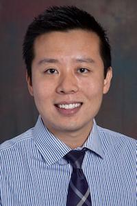
Ranliang Hu, MD
Assistant Professor
Emory University
© 2026 Medality. All Rights Reserved.
