Training Collections
Library Memberships
On-demand course library with video lectures, expert case reviews, and more
Fellowship Certificate™ Programs
Practice-focused training programs designed to help you gain experience in a specific subspecialty area.
Ultimate Learning Pass
Unlock access to our full Course Library and all self-paced Fellowships.
Continuing Medical Education (State CME)
Complete all of your state CME requirements in one convenient place.
Noon Conference (Free)
Get access to free live lectures, every week, from top radiologists.
Case of the Week (Free)
Get a free weekly case delivered right to your inbox.
Case Crunch: Rapid Case Review (Free)
Register for free live board reviews.
Dr. Resnick's MSK Conference
Learn directly from the MSK Master himself.
Lower Extremities MRI Conference
Musculoskeletal Imaging
For Training Programs
Supplement your training program with case-based learning for residents, registrars, fellows, and more.
For Private Practices
Upskill in high growth, advanced imaging areas.
Compliance
NewTrack, fulfill, and report on all your radiologists' credentialing and licensing requirements.
Emergency Call Prep
Prepare trainees to be on call for the emergency department with this specialized training series.
Training Collections
Library Memberships
On-demand course library with video lectures, expert case reviews, and more
Fellowship Certificate™ Programs
Practice-focused training programs designed to help you gain experience in a specific subspecialty area.
Ultimate Learning Pass
Unlock access to our full Course Library and all self-paced Fellowships.
Continuing Medical Education (State CME)
Complete all of your state CME requirements in one convenient place.
Noon Conference (Free)
Get access to free live lectures, every week, from top radiologists.
Case of the Week (Free)
Get a free weekly case delivered right to your inbox.
Case Crunch: Rapid Case Review (Free)
Register for free live board reviews.
Dr. Resnick's MSK Conference
Learn directly from the MSK Master himself.
Lower Extremities MRI Conference
Musculoskeletal Imaging
For Training Programs
Supplement your training program with case-based learning for residents, registrars, fellows, and more.
For Private Practices
Upskill in high growth, advanced imaging areas.
Compliance
NewTrack, fulfill, and report on all your radiologists' credentialing and licensing requirements.
Emergency Call Prep
Prepare trainees to be on call for the emergency department with this specialized training series.
Lesson
Sample Case Review
1 topic, 3 min. of video
Lesson
Breast Calcifications Expert Case Reviews
13 topics, 37 min. of video
Management of Breast Calcifications - Case 2
4 m.Management of Breast Calcifications - Case 3
4 m.Management of Breast Calcifications - Case 4
5 m.Management of Breast Calcifications - Case 5
2 m.Management of Breast Calcifications - Case 6
2 m.Management of Breast Calcifications - Case 7
6 m.Management of Breast Calcifications - Case 8
3 m.Management of Breast Calcifications - Case 9
3 m.Management of Breast Calcifications - Case 10
2 m.Management of Breast Calcifications - Case 11
5 m.Management of Breast Calcifications - Case 12
2 m.Management of Breast Calcifications - Case 13
4 m.Management of Breast Calcifications - Case 14
2 m.0:00
This is a case number 10 where a 70
0:03
year old was called back from screening.
0:06
So on the spot images you could see
0:09
there's a solitary math.
0:12
With indistinct margins you see the margins
0:15
are in distinct here and here and you
0:18
could see there's core scalifications again.
0:21
All of these worrisome
0:24
features should prompt biopsy.
0:27
And in this case, it was
0:30
invasive cancer with metaplastic features.
0:37
I just want to show you a companion case with masks with
0:40
calcification here. You see a 60 year old with bilateral palpable
0:43
masses here in contrast
0:46
to the priorities that I shown there are multiple
0:49
oval masses with circumscribed margin
0:52
and some of
0:55
this containing course pop-like calcifications.
0:58
These are in the Looting fibroidinomas and
1:01
it's by rats too. So here you could see
1:04
there's different phases of inverloading fibroid enormous.
1:07
So here you just see the mass and then
1:10
here you could see the mass.
1:13
It starts with peripheral calcification and
1:16
in the Final Phase, you just see the
1:19
popcorn calcification and you don't see the
1:22
underlying Mass again this when you
1:25
have multiple circumscribed masses you want
1:28
to it's usually a sign of benignity
1:31
whereas um, when I when I show
1:34
you back on the prior study always beware
1:37
of solitary Mass with a non-circumscribed
1:40
margin associated with calcifications.
Management of Breast Calcifications - Case 10
HIDEInteractive Transcript
0:00
This is a case number 10 where a 70
0:03
year old was called back from screening.
0:06
So on the spot images you could see
0:09
there's a solitary math.
0:12
With indistinct margins you see the margins
0:15
are in distinct here and here and you
0:18
could see there's core scalifications again.
0:21
All of these worrisome
0:24
features should prompt biopsy.
0:27
And in this case, it was
0:30
invasive cancer with metaplastic features.
0:37
I just want to show you a companion case with masks with
0:40
calcification here. You see a 60 year old with bilateral palpable
0:43
masses here in contrast
0:46
to the priorities that I shown there are multiple
0:49
oval masses with circumscribed margin
0:52
and some of
0:55
this containing course pop-like calcifications.
0:58
These are in the Looting fibroidinomas and
1:01
it's by rats too. So here you could see
1:04
there's different phases of inverloading fibroid enormous.
1:07
So here you just see the mass and then
1:10
here you could see the mass.
1:13
It starts with peripheral calcification and
1:16
in the Final Phase, you just see the
1:19
popcorn calcification and you don't see the
1:22
underlying Mass again this when you
1:25
have multiple circumscribed masses you want
1:28
to it's usually a sign of benignity
1:31
whereas um, when I when I show
1:34
you back on the prior study always beware
1:37
of solitary Mass with a non-circumscribed
1:40
margin associated with calcifications.
Report
Faculty
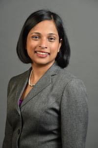
Jean Kunjummen, DO
Associate Professor, Department of Radiology and Imaging Sciences,
Emory School of Medicine
Tags
Screening
Mammography
Breast
© 2026 Medality. All Rights Reserved.
