Training Collections
Library Memberships
On-demand course library with video lectures, expert case reviews, and more
Fellowship Certificate™ Programs
Practice-focused training programs designed to help you gain experience in a specific subspecialty area.
Ultimate Learning Pass
Unlock access to our full Course Library and all self-paced Fellowships.
Continuing Medical Education (State CME)
Complete all of your state CME requirements in one convenient place.
Noon Conference (Free)
Get access to free live lectures, every week, from top radiologists.
Case of the Week (Free)
Get a free weekly case delivered right to your inbox.
Case Crunch: Rapid Case Review (Free)
Register for free live board reviews.
Dr. Resnick's MSK Conference
Learn directly from the MSK Master himself.
Lower Extremities MRI Conference
Musculoskeletal Imaging
Emergency Imaging
Emergency GI & Abdominal Radiology FellowshipNEW!
PET Imaging
Pediatric Imaging
For Training Programs
Supplement your training program with case-based learning for residents, registrars, fellows, and more.
For Private Practices
Upskill in high growth, advanced imaging areas.
Compliance
NewTrack, fulfill, and report on all your radiologists' credentialing and licensing requirements.
Emergency Call Prep
Prepare trainees to be on call for the emergency department with this specialized training series.
Training Collections
Library Memberships
On-demand course library with video lectures, expert case reviews, and more
Fellowship Certificate™ Programs
Practice-focused training programs designed to help you gain experience in a specific subspecialty area.
Ultimate Learning Pass
Unlock access to our full Course Library and all self-paced Fellowships.
Continuing Medical Education (State CME)
Complete all of your state CME requirements in one convenient place.
Noon Conference (Free)
Get access to free live lectures, every week, from top radiologists.
Case of the Week (Free)
Get a free weekly case delivered right to your inbox.
Case Crunch: Rapid Case Review (Free)
Register for free live board reviews.
Dr. Resnick's MSK Conference
Learn directly from the MSK Master himself.
Lower Extremities MRI Conference
Musculoskeletal Imaging
Emergency Imaging
Emergency GI & Abdominal Radiology FellowshipNEW!
PET Imaging
Pediatric Imaging
For Training Programs
Supplement your training program with case-based learning for residents, registrars, fellows, and more.
For Private Practices
Upskill in high growth, advanced imaging areas.
Compliance
NewTrack, fulfill, and report on all your radiologists' credentialing and licensing requirements.
Emergency Call Prep
Prepare trainees to be on call for the emergency department with this specialized training series.
Lesson
Sample Lesson - Meningiomas
1 topic, 5 min. of video
Lesson
Meningiomas
15 topics, 57 min. of video
Assessing Lesion Position
4 m.Intra-axial vs. Extra-axial Lesions
3 m.Typical Locations of Meningiomas
3 m.Defining Meningioma
2 m.Meningioma Appearance on MRI
6 m.Vascular Composition
3 m.Meningioma vs. Schwannoma
5 m.Meningiomas in the Posterior Fossa
5 m.Optic Nerve Sheath Meningioma
4 m.Vasogenic Edema
3 m.Suprasellar Meningioma
7 m.Optic Nerve Meningioma
6 m.Juxtasellar Meningioma
5 m.Meningiomatosis
5 m.Hemangiopericytoma
6 m.Lesson
Non-glial Tumors
13 topics, 45 min. of video
Hemangioblastoma Part 1
6 m.Hemangioblastoma Part 2
3 m.Spinal Hemangioblastoma
4 m.Recurrent Hemangioblastoma
3 m.VHL Renal Lesions Part 1
3 m.VHL Renal Lesions Part 2
3 m.Hemangioblastoma with Mass Effect Part 1
3 m.Hemangioblastoma with Mass Effect Part 2
3 m.Endolymphatic Sac Tumor
3 m.Central Neuro, Pediatricscytoma
6 m.Lhermitte Duclos Disease
6 m.Epidermoid Cyst
4 m.Rhabdomyosarcoma
4 m.0:00
I'm here with my brilliant young colleague,
0:02
Dr. Ben Laser.
0:03
We're talking about the vascular makeup of meningiomas.
0:06
This is a 43-year-old man with a huge falcine meningioma
0:09
with left-sided symptoms from compression.
0:12
The meningioma is on the right side.
0:14
It's a huge falcine meningioma seen on T1,
0:17
T1 C+ and T1 coronal.
0:19
So this is an axial, this is a coronal,
0:21
T1 C+.
0:23
And this lesion demonstrates some rather intense
0:26
enhancement with some dural enhancement.
0:28
We talked previously about the mother-in-law sign.
0:31
Both of us are successfully married.
0:33
Mother-in-laws are terrific.
0:35
They come early to help you out with the kids,
0:37
but they also stay late to babysit
0:39
to help you out with the kids.
0:40
Some people say that the enhancement of a meningioma
0:43
is intense, kind of like your mother-in-law.
0:46
But the question that I have is why?
0:48
And you know there are some normal areas of
0:50
the brain that normally enhance,
0:52
like the dura and the choroid plexus,
0:55
and the area postrema and the pineal gland.
0:59
So why do meningiomas have this intense enhancement,
1:03
not unlike some normal structures?
1:05
So these normal structures that Dr. Pomeranz
1:07
has just described, all have a type
1:09
of fenestration in their capillaries.
1:11
There's three different types of capillaries.
1:13
You have the continuous capillary,
1:14
you have the sinusoidal capillaries,
1:16
and then you have the fenestrated capillaries.
1:18
The fenestrated capillaries, in all these areas, take up
1:21
enhancement and allow the blood that has the contrast
1:26
to enhance these regions.
1:26
So, they're more leaky.
1:27
So they're more leaky.
1:28
Essentially, the vessels are leaky,
1:30
and that's why the meningioma enhances
1:33
so rapidly and avidly.
1:35
So, basically what happens is
1:36
these normal blood vessels
1:38
that are different, the subtype of capillary
1:40
known as autochthonous blood supply in these areas become
1:44
too proliferative in the meningioma and allow very rapid
1:48
leakage of gadolinium or the contrast
1:50
agent into the lesion,
1:52
producing a mass that exhibits the mother-in-law sign,
1:55
comes early, stays late.
1:57
It's intense.
1:58
Let's move on, shall we?
Vascular Composition
HIDEInteractive Transcript
0:00
I'm here with my brilliant young colleague,
0:02
Dr. Ben Laser.
0:03
We're talking about the vascular makeup of meningiomas.
0:06
This is a 43-year-old man with a huge falcine meningioma
0:09
with left-sided symptoms from compression.
0:12
The meningioma is on the right side.
0:14
It's a huge falcine meningioma seen on T1,
0:17
T1 C+ and T1 coronal.
0:19
So this is an axial, this is a coronal,
0:21
T1 C+.
0:23
And this lesion demonstrates some rather intense
0:26
enhancement with some dural enhancement.
0:28
We talked previously about the mother-in-law sign.
0:31
Both of us are successfully married.
0:33
Mother-in-laws are terrific.
0:35
They come early to help you out with the kids,
0:37
but they also stay late to babysit
0:39
to help you out with the kids.
0:40
Some people say that the enhancement of a meningioma
0:43
is intense, kind of like your mother-in-law.
0:46
But the question that I have is why?
0:48
And you know there are some normal areas of
0:50
the brain that normally enhance,
0:52
like the dura and the choroid plexus,
0:55
and the area postrema and the pineal gland.
0:59
So why do meningiomas have this intense enhancement,
1:03
not unlike some normal structures?
1:05
So these normal structures that Dr. Pomeranz
1:07
has just described, all have a type
1:09
of fenestration in their capillaries.
1:11
There's three different types of capillaries.
1:13
You have the continuous capillary,
1:14
you have the sinusoidal capillaries,
1:16
and then you have the fenestrated capillaries.
1:18
The fenestrated capillaries, in all these areas, take up
1:21
enhancement and allow the blood that has the contrast
1:26
to enhance these regions.
1:26
So, they're more leaky.
1:27
So they're more leaky.
1:28
Essentially, the vessels are leaky,
1:30
and that's why the meningioma enhances
1:33
so rapidly and avidly.
1:35
So, basically what happens is
1:36
these normal blood vessels
1:38
that are different, the subtype of capillary
1:40
known as autochthonous blood supply in these areas become
1:44
too proliferative in the meningioma and allow very rapid
1:48
leakage of gadolinium or the contrast
1:50
agent into the lesion,
1:52
producing a mass that exhibits the mother-in-law sign,
1:55
comes early, stays late.
1:57
It's intense.
1:58
Let's move on, shall we?
Report
Description
Faculty
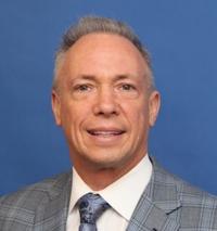
Stephen J Pomeranz, MD
Chief Medical Officer, ProScan Imaging. Founder, MRI Online
ProScan Imaging
Tags
Pediatrics
Neuroradiology
Neoplastic
MRI
Brain
© 2026 Medality. All Rights Reserved.
