- Encephalomalacia is the end result of liquefactive necrosis in the brain, commonly caused by infarction, hemorrhage, or trauma.
- It can occur in any age group and location, with the anteroinferior frontal and temporal lobes often affected after trauma.
- Lesions may be asymptomatic or lead to neurologic deficits or seizures, depending on their location.
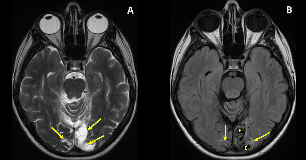
- On imaging, it shows CSF-like signal, volume loss, and negative mass effect
What Is a Encephalomalacia?
Encephalomalacia, which literally means “softening of the brain,” is a radiologic finding that often raises critical diagnostic and prognostic questions. Whether you’re a radiology resident honing your interpretive skills or a seasoned radiologist reviewing complex neuroimaging, understanding encephalomalacia is essential for clinical accuracy and confident reporting.
Definition and Pathophysiology
Encephalomalacia is a nonspecific term used to describe the end result of liquefactive necrosis of brain parenchyma. It can be focal or diffuse, and it is not limited by age, it can be observed in adults, children, and even in utero. This softening represents irreversible damage to brain tissue, which is eventually replaced by cerebrospinal fluid (CSF).
The condition can occur in any region of the brain, but after trauma, characteristic locations include the anteroinferior frontal lobes and temporal lobes.
Feature by feature
| Feature | Encephalomalacia (chronic) | Why it matters |
|---|---|---|
| Volume loss | The nearby ventricle enlarges and sulci widen toward the lesion (ex vacuo dilatation). There is no mass effect. | Acute infarct, edema and tumor push structures away. Encephalomalacia pulls them in. |
| T1 | Dark, often matching CSF in the cystic center. | Confirms the tissue is gone, not just swollen. |
| T2 | Bright, matching CSF. | On its own this doesn’t help much, because edema, tumor and acute infarct are also T2 bright. |
| FLAIR | The cystic center suppresses and turns dark like CSF. A bright rim of gliosis surrounds it. | This is the key sequence. Acute and subacute lesions stay bright on FLAIR. They don’t suppress. |
| DWI / ADC | No restricted diffusion. DWI is dark and ADC is bright, because water moves freely in the cavity. | Separates it from an acute infarct, which is bright on DWI and dark on ADC. |
| SWI / GRE | May show dark blooming from hemosiderin. | Points to old hemorrhage or trauma as the cause. |
| Contrast | No enhancement. | Enhancement suggests a subacute infarct (about 1 to 8 weeks), tumor or infection. |
What Causes Encephalomalacia?
The most common causes of encephalomalacia include:
- Ischemic stroke (chronic infarct)
- Traumatic brain injury
- Intracranial hemorrhage
- Infections such as encephalitis or abscess
- Surgical resection or radiation necrosis
In pediatric patients, it may also result from perinatal hypoxic-ischemic injury or congenital anomalies.
What’s the Clinical Significance of Encephalomalacia?
Encephalomalacia can be an incidental finding or correlate directly with neurologic symptoms such as:
- Seizures
- Cognitive decline
- Motor or sensory deficits
- Behavioral changes
The location and extent of the lesion often influence clinical presentation and guide management decisions.
How Should Radiologists Report Encephalomalacia?
When encephalomalacia is identified, radiologists should describe:
- Location and size of the lesion
- Associated findings such as gliosis, hemosiderin deposition, or midline shift
- Possible etiology, especially if signs of prior infarct, trauma, or hemorrhage are present
- Impact on adjacent structures (e.g., mass effect, ventricular distortion)
This context helps referring physicians determine if further workup, neurologic referral, or supportive care is required.
Quick checklist
- Does it pull or push? Pulling the ventricle or sulci toward it points to tissue loss.
- FLAIR: Does the center turn dark with a bright rim? That is classic encephalomalacia with gliosis.
- DWI/ADC: Is restricted diffusion absent? If so, the lesion isn’t acute.
- Contrast: Is there no enhancement? That suggests a chronic, inactive lesion.
- SWI: Is there blooming? That suggests old blood, from trauma or a prior hemorrhage.
- Distribution: The pattern hints at the cause. A vascular territory suggests infarct. The frontal and temporal poles suggest contusion. A lesion next to a surgical site suggests a postoperative change.
Learn More and Review Diagnostic Cases
At Medality, we help radiologists and trainees sharpen their diagnostic accuracy with real-world cases and expert-led reviews. Our Encephalomalacia diagnosis page includes:
- Clinical imaging examples
- Case-based learning
- Tips for identifying encephalomalacia
And our Brain & Spine Fellowship Certificate™ program is a case-based learning approach for further specialization in neuroradiology.