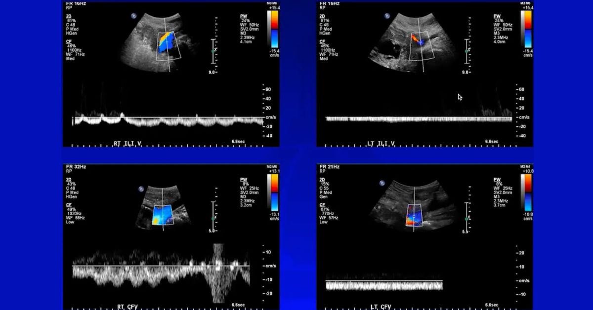
When evaluating lower extremity swelling, vascular ultrasound is often the first tool used to detect venous obstruction or thrombosis. But not all findings are straightforward. Subtle waveform changes can easily be overlooked without the right experience.
In this free case review from our Peripheral Vascular Ultrasound Course, you’ll walk through a real example of dampened flow in the left external iliac vein, and learn how to recognize and interpret a common yet critical finding in vascular imaging.
About the Peripheral Vascular Ultrasound Course
This free case is just one of many inside our Peripheral Vascular Ultrasound Course (within Medality's Vascular Imaging library). In this course, Dr. Sheila Sheth:
- Reviews technique and ultrasound abnormalities in lower and upper extremity vascular ultrasound
- Helps you understand the implications of abnormal waveforms
- Walks through a series of interesting and challenging peripheral venous and arterial cases
If you found this case helpful, the full Peripheral Vascular Ultrasound Course is packed with real-world cases just like this one, all designed to help you interpret faster, with more confidence.
Start learning in just 5 minutes a day and earn CME along the way.


